Elevating throughput without compromising quality

The first multi-well HD-MEA platform capable of running 24 & 96 well HD-MEAs with simultaneous recording from all wells and all electrodes.

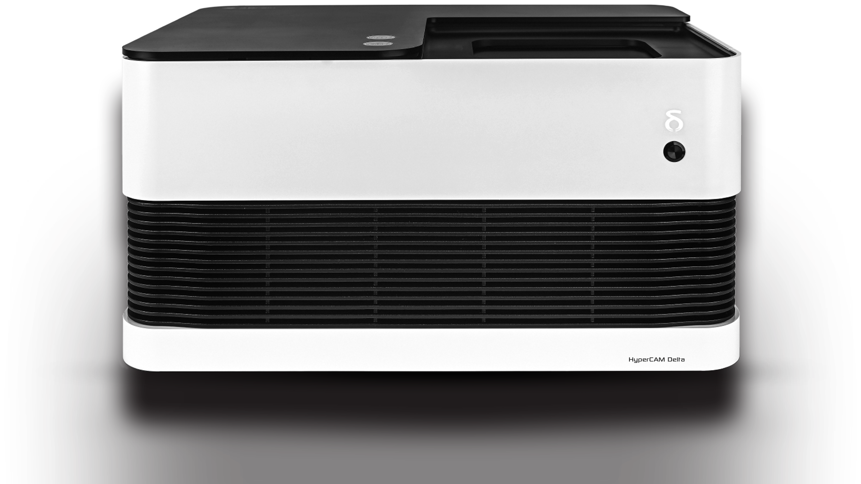
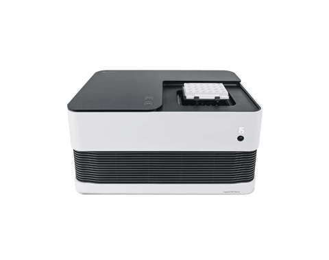
Front view of a HyperCAM Delta projector with a white and black rectangular design.

Key Benefits

Automation ready
Simultaneous Recording
High-Throughput Research

Simultaneous recording of ALL wells, and ALL electrodes

Rat Primary Cortical Neuronal Culture 24 DIV

Overview

Built with progression in mind, anticipating future demands.

The system is designed for scalability, supporting both 24 & 96 well HD-MEA format & robotic automation. The HyperCAM Delta allows you to further expand your research capacity as needed, ensuring you can maximize data collection and streamline your research processes, both now and in the future.

Obtain more data, more efficiently without compromising quality.

The HyperCAM Delta is a truly unique multi-well HD-MEA platform. With the highest number of simultaneous recording wells and electrodes available on the market, the system enables you to generate high-quality, high-resolution data with exceptional statistical strength. Reducing the need for excessive replicates, accelerating results, and enhancing cost-efficiency throughout your workflow.

High throughput, high accuracy functional screening of your electrogenic cells.

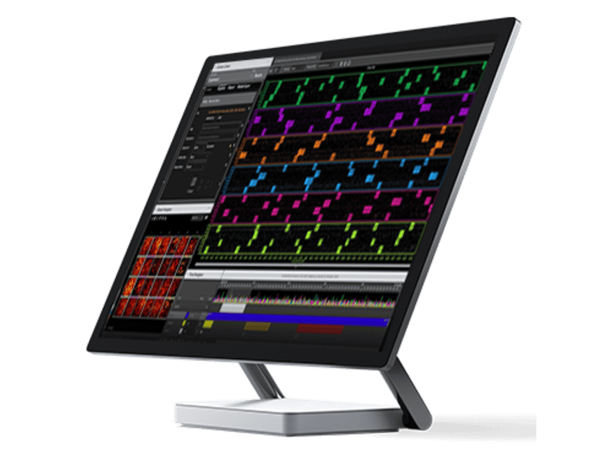
When paired with the revolutionary CorePlate™ 24W or CorePlate™ 96W, the HyperCAM Delta delivers truly unmatched HD‑MEA performance, recording from ALL wells and ALL electrodes simultaneously, the system enables highly efficient acquisition of large‑scale, high‑value datasets. Built with an integrated environmental chamber and full automation compatibility, the HyperCAM Delta further streamlines your workflow from start to finish. When combined with BrainWave software, navigating, visualizing, and analysing these extensive datasets becomes both intuitive and efficient. The combination of high‑density data capture, automation readiness, and seamless analysis makes the HyperCAM Delta the ideal platform for advanced multi‑well HD‑MEA research.

Compatibility

High density multiwell sensors for high definition results

HyperCAM Alpha is a CorePlate™ powered 6 well platform that handles parallel experiments with neuronal cultures, organoids or brain slices. 4096 fully bidirectional electrodes (recording/stimulating) per well allow real time characterization of neuronal diseases, mutations and cell effectors with a high statistical precision.

The CorePlate 24W 16/50.

The inaugural 24 well HD-MEA on the market offering simultaneous recording across all wells and all 24,576 electrodes (1,024 electrodes per well), enabling unparalleled data density in a multi-well format.

Logo of CorePlate™ 96W
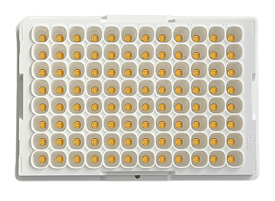
White 96-well microplate with 8 rows and 12 columns of wells, each containing a small yellow square in the center.

The first 96 well HD-MEA, available to order in 2026, featuring simultaneous recording across all wells and all 24,576 electrodes (256 electrodes per well), enabling true high-throughput electrophysiology.

Overview

Built with progression in mind, anticipating future demands.

The system is designed for scalability, supporting both 24 & 96 well HD-MEA format & robotic automation. The HyperCAM Delta allows you to further expand your research capacity as needed, ensuring you can maximize data collection and streamline your research processes, both now and in the future. 

Obtain more data, more efficiently without compromising quality.

The HyperCAM Delta is a truly unique multi-well HD-MEA platform. With the highest number of simultaneous recording wells and electrodes available on the market, the system enables you to generate high-quality, high-resolution data with exceptional statistical strength. Reducing the need for excessive replicates, accelerating results, and enhancing cost-efficiency throughout your workflow.

High throughput, high accuracy functional screening of your electrogenic cells.

When paired with the revolutionary CorePlate™ 24W or CorePlate™ 96W, the HyperCAM Delta delivers truly unmatched HD‑MEA performance, recording from ALL wells and ALL electrodes simultaneously, the system enables highly efficient acquisition of large‑scale, high‑value datasets. Built with an integrated environmental chamber and full automation compatibility, the HyperCAM Delta further streamlines your workflow from start to finish. When combined with BrainWave software, navigating, visualizing, and analysing these extensive datasets becomes both intuitive and efficient. The combination of high‑density data capture, automation readiness, and seamless analysis makes the HyperCAM Delta the ideal platform for advanced multi‑well HD‑MEA research.

Compatibility

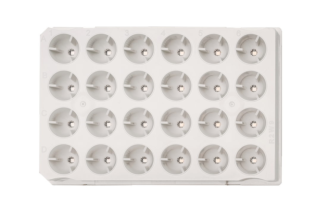
Empty 24-well cell culture plate with grid labeling of rows A to D and columns 1 to 6.

The inaugural 24 well HD-MEA on the market offering simultaneous recording across all wells and all 24,576 electrodes (1,024 electrodes per well), enabling unparalleled data density in a multi-well format.

The first 96 well HD-MEA, available to order in 2026, featuring simultaneous recording across all wells and all 24,576 electrodes (256 electrodes per well), enabling true high-throughput electrophysiology.

Applications
The HyperCAM Delta is designed for a wide range of applications.
Record from Neuronal and Cardiac
An icon for primiary cultures.
Primary cultures 
An icon for HiPSC derived cultures.
HiPSC derived cultures
An icon for Spheroids.
Spheroids
An icon for Organoids.
Organoids
To perform
An icon for disease model characterization.
Toxicology assays
An icon for disease model characterization.
Disease model characterization
An icon showing drug screening.
Drug screening
An icon showing safety pharmacoogy.
Safety pharmacology
Data examples
Primary Neuronal Culture
Hippocampal Culture 17 DIV
This image represents a visual data analysis of neural activity captured via a multi-electrode array (MEA) system, used in electrophysiology research. The image contains four main sections, each labeled (a, b, c, d) and connected to a multi-well plate on the left side of the image.
Rat Hippocampal Culture 17 DIV showing a. Activity map showing Mean Firing Rate (spikes / s). b. Raw signal map (change in µV in a 100ms window).
c. Raw signal trace showing burst activity, with spikes identified below (black vertical lines) and a detected burst (horizontal blue line). d. Raster Plot showing synchronized activity throughout a Rat Hippocampal Culture at 17 DIV.
Primary Neuronal Culture
Hippocampal Culture 17 DIV
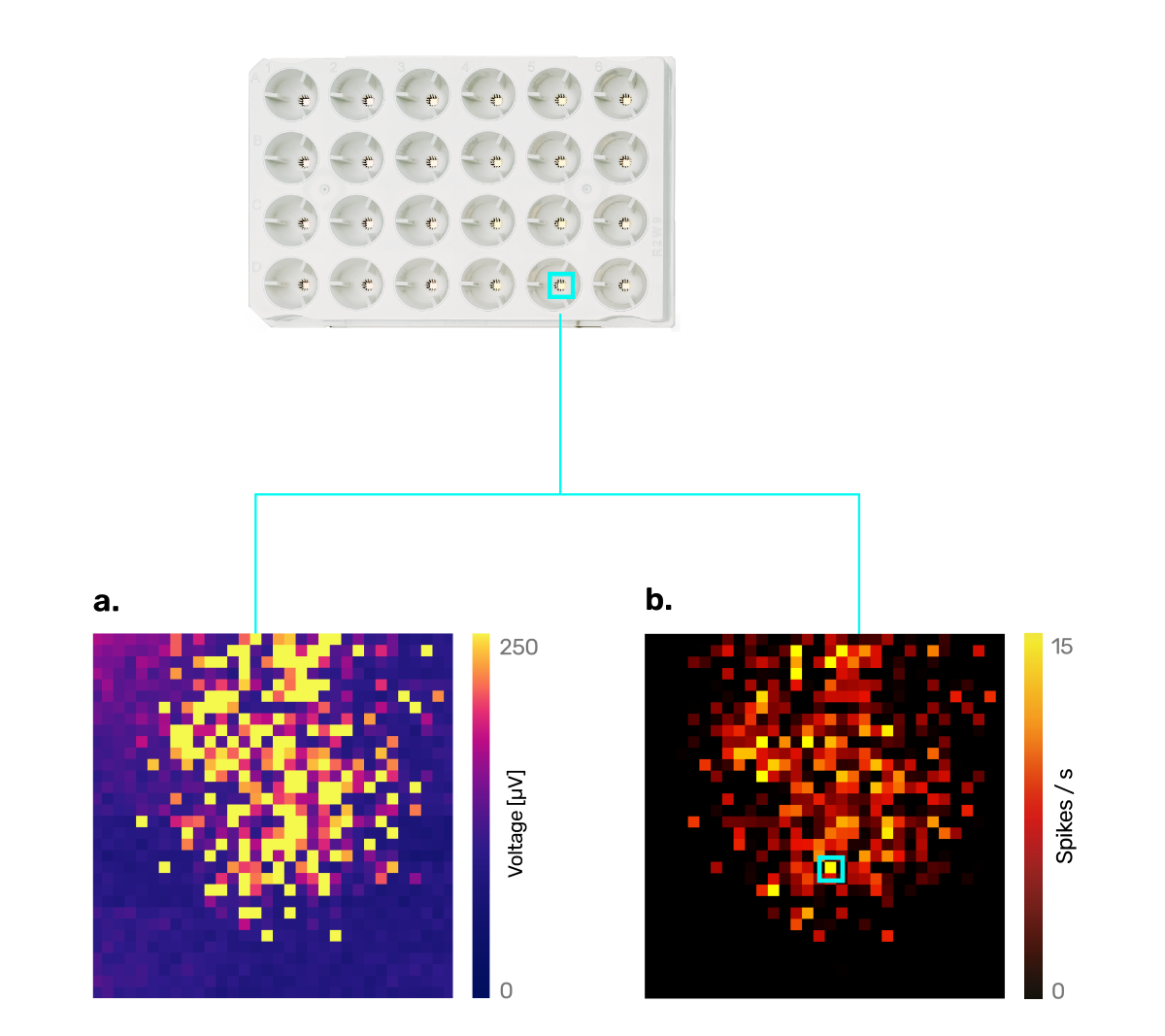
Top view of a 4x6 grid electrode array with two heatmaps below; left heatmap shows voltage distribution in microvolts, right heatmap shows spike rates in spikes per second, both highlighting data from the bottom right electrode.
Rat Hippocampal Culture 17 DIV showing  a. Raw signal map (change in µV in a 100ms window).  b. Activity map showing Mean Firing Rate (spikes / s),
Graph titled Raw Traces showing voltage in microvolts with fluctuating black line peaks around 02:39.40, ranging from -500 to 500.
Raw signal trace showing burst activity, with spikes identified below (white vertical lines) and a detected burst (horizontal yellow line).
Raster plot displaying electrode signals over time with electrodes on the y-axis and time in seconds on the x-axis, showing multiple vertical spike lines.
Raster Plot showing synchronized activity throughout a Rat Hippocampal Culture at 17 DIV.
Hippocampal Culture 11 DIV
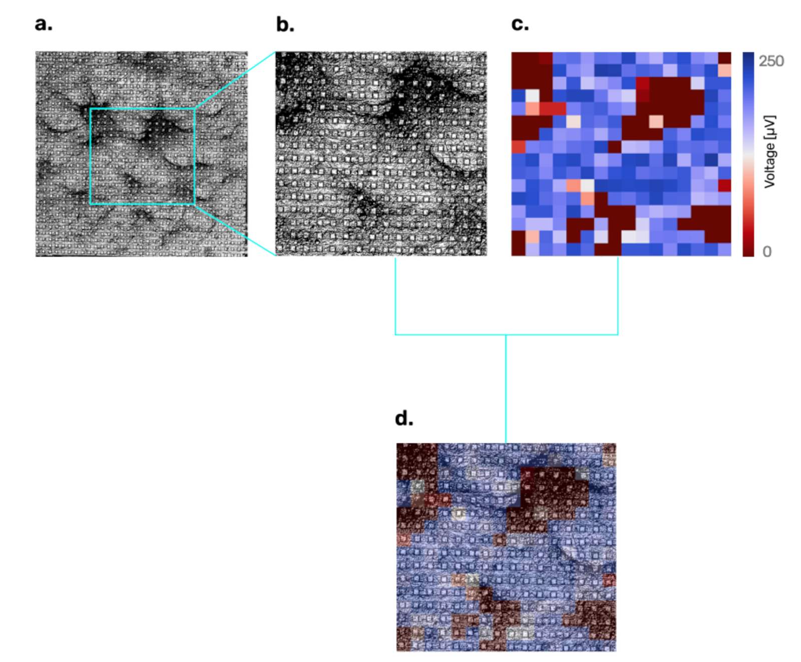
Four connected images: a black and white grayscale micrograph showing circular pores, magnified inset of the same micrograph highlighting pore patterns, a heatmap with blue and red colors indicating a frequency scale from 0 to 250, and a combined overlay of the heatmap on the micrograph.

a. Rat Hippocampal Culture at 11 DIV over CorePlateTM. b. Magnification of a central area of the culture. c. Activity map measured by changes in µV in a 100ms window. d. Overlay of the activity map over the culture image.

Spheroids
Human Derived Cortical Spheroids 45 DIV

Spiking activity map (spikes / s) and associated raw trace of human derived cortical spheroids 45 DIV

Cardiomyocyte Culture
Cardiomyocyte Culture 9 DIV
Raw trace from a cardiomyocyte culture 9 DIV displaying a clear initial QRS complex followed by a T wave.
DATA EXAMPLES
Primary Neuronal Culture
Hippocampal Culture 17 DIV
Image showing a 24-electrode grid with highlighted electrode connected to two heatmaps: a) voltage map with yellow and purple scale, b) spikes per second map with red, orange, and black scale.
Rat Hippocampal Culture 17 DIV showing  a. Raw signal map (change in µV in a 100ms window).  b. Activity map showing Mean Firing Rate (spikes / s)
Graph titled 'Raw Traces' showing voltage in microvolts over time with a spike in voltage around 02:39.40.
Raw signal trace showing burst activity, with spikes identified below (white vertical lines) and a detected burst (horizontal yellow line).
Raster plot showing neural spike times with electrodes on the vertical axis and time in seconds on the horizontal axis.
Raster Plot showing synchronized activity throughout a Rat Hippocampal Culture at 17 DIV.
Hippocampal Culture 11 DIV
Four-panel scientific visualization showing tissue imaging: a) wide view of tissue section with cellular features; b) zoomed-in detail of a selected tissue area showing detailed cellular structures; c) heatmap representing spatial protein expression levels ranging from 0 to 250; d) combined overlay of tissue image with protein expression heatmap highlighting spatial patterns.
a. Rat Hippocampal Culture at 11 DIV over CorePlateTM. b.Magnification of a central area of the culture. c.Activity map measured by changes in µV in a 100ms window. d. Overlay of the activity map over the culture image.
Spheroids
Human Derived Cortical Spheroids 45 DIV

Spiking activity map (spikes / s) and associated raw trace of human derived cortical spheroids 45 DIV

Cardiomyocyte Culture
Cardiomyocyte Culture 9 DIV
Raw trace from a cardiomyocyte culture 9 DIV displaying a clear initial QRS complex followed by a T wave.

Tech specs

Signal Acquisition Module

Computational core

Multi-FPGA parallel processing with 5x Intel®’s Arria® 10 FPGA 13Gbps, 4GB DDR4, and ARM® dual-core Cortex™-A9 1.5 GHz

HD-MEA Compatibility

CorePlate™ 24W 16/50 (requires 24w/25ke Module)  
CorePlate™ 96W 16/50 (requires 96w/25ke Module)

Max number of simultaneous recording wells

Depending on the installed module:
24 (with 24w/25ke Module)
96 (with 96w/25ke Module)

Max number of simultaneous recording electrodes

Depending on the installed module:
24,576 @ 10 kHz (with 24w/25ke Module)
24,575 @ 10 kHz (with 96w/25ke Module)

Data resolution

11 bit

Electrode subset recording

Yes, predefined regions

Max sampling frequency

10 kHz

Physical specs

Power supply requirements

110-240 VAC, 50/60 Hz, 2 A

Body material

ABS/PC blend and anodized aluminum for the envrionmental chamber

Dimensions (W x D x H)

530 x 360 x 289 mm (20.9 x 14.2 x 11.4 in)

Weight

24.0 kg (53.0 lbs)

Gross weight (with box, accessories...)

33.0 kg (72.8 lbs)

CorePlate™ Multi-wells

Discover more

BrainWave 6

Discover more

Resources

Here you can find useful resources for your HyperCAM Delta

HyperCAM Delta Brochure
Can't find what you're looking for? No worries! Reach out to us—our team is here and happy to help.
Contact Us