Electrophysiological Signals & Microelectrode Array Principles

Basic principles

What is an electrophysiological signal?

A cellular membrane is a semi permeable lipid bilayer acting as a capacitor that accumulates positive and negative ions on each side of the membrane. At rest, cells like neurons accumulate negatively charged ions in the intracellular compartment. Because the membranes are not permeable to ions, the asymmetry of charges on each side of the membrane creates a difference of electrical potential (or voltage) called the membrane potential. This membrane potential can be measured with intracellular techniques such as sharp electrodes or patch clamp.

Neuronal activity, by opening specific ion channels, generates currents due to the ionic movements driven by their electrochemical gradient. Because of the high resistance of the contact between the neuron and the electrode, these small currents generate a non-neglectable voltage (ohm law). The quality of the recorded signal depends greatly on the proximity and the nature of the contact between the electrode and the cell: the closer is the cell or the better is the contact (the higher is the resistance), the better is the signal recorded. These voltage changes can be measured from inside the cell with intracellular electrodes, or at a distance with extracellular electrodes positioned almost in contact with the cell.

What is an electrophysiological signal? - 3Brain

An extracellular electrode can record the extracellular field potential generated by the action potentials of one or more neurons. It reflects the force (or electrical field) exerted on ions in a conductive medium like the extracellular compartment (Graziane and Dong, 2016). It is then an indirect measurement of the ionic movements that corresponds in principle to the first derivative of the signal sensed in an intracellular configuration (Henze et al, 2000). Recorded signals can reflect the fast spike activity of individual neurons (events in the order of 0.5-2 msec with hundreds of µV peak amplitude) or the slower superposition of action potentials and synaptic potentials (Spira et al, 2013) resulting in waves (>10 ms duration) of high amplitude (typically from hundreds of µV to 1-2 mV).

Traditional intracellular recording techniques such as patch clamp provide a better signal quality compared to conventional extracellular approaches, but they have severe technical limitations in scaling up the number of sensing sites and therefore cannot efficiently decipher neural network function in health and disease. This results in the impossibility to describe the physio-pathological neuronal activity at a level where the neuron stops acting as a single unit and starts working in cooperation with other cells, providing the parallelized, integrative and massively multisensorial processing that is a unique signature of the brain. Therefore, developing approaches to increase the number of recorded neurons has been a major neurotechnological challenge of the last decades (Maccione et al, 2015).

Microelectrode array from 3Brain

(adapted from Imfeld et al, 2008) - 3Brain

(adapted from Imfeld et al, 2008)

The active electronics start conditioning the recorded signals directly on the chip. Every single electrode has an active, intelligent technology behind it. Amplifiers are located right below each electrode, allowing enhancement of the signal at the source (right underneath the neurons) without amplifying any additional noise, and preventing signal attenuation during the propagation of the signal. This is how 3Brain’s HD-MEA can reach an outstanding Signal to Noise Ratio (SNR).

The pixels are square electrodes of 21 μm, arranged in a 64 by 64 layout with an electrode pitch comprised between 42 µm and 81 µm. Signals can be recorded at a sampling rate of 18 to 64 KHz for each pixel.

What is a microelectrode array? How does a microelectrode array work?

(figure adapted from Berdondini et al, 2009) - 3Brain

(figure adapted from Berdondini et al, 2009)

3Brain’s CMOS-MEA chip is based on the Active Pixel Sensor (APS) concept commonly used in high-speed digital cameras, in which the original in-pixel circuitry was entirely redesigned to record small extracellular voltage variations resulting from cellular activity instead of detecting light changes. It acts as a sort of video camera: the signal is collected as frames, where each point represents the instant extracellular voltage value for each pixel. It is then possible to reconstruct the signal into a video of the neuronal activity by following a single pixel over time. This video is color-coded depending of the level of activity. The signal can also be visualized as single electrode traces over time by selecting one or many pixels. 3Brain revolutionized the field by introducing the concept of “functional imaging” in the microelectrode array world, providing for the first time movies of the electrical activity generated by large portions of neuronal tissues (Ferrea et al, 2012).

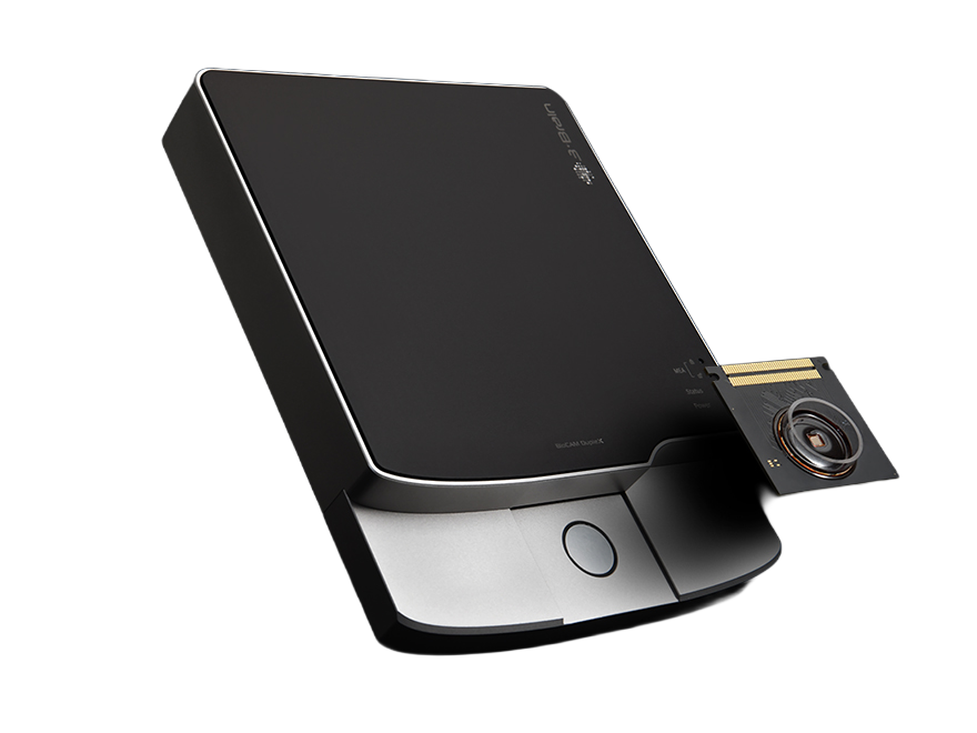
3Brain’s current CorePlate™ Technology relies on an outstanding chip unique in the MEA world which features thousands of extracellular electrodes per well that can be recorded simultaneously or used to release an electrical stimulation.

The previous single-well generation, known as the HD-MEA (High Density Microelectrode or Multielectrode Array), had already set the gold standard with 4096 simultaneous recording/stimulating electrodes.

Advantages

3Brain’s single-well and multi-well CorePlate™ powered, based on high density microelectrode array technology, allow studying the physiological and pathological functional activity indifferent models such as cell culture (primary or stem cell derived), brain slices (acute or organotypic), retina or brain organoids.

They are versatile tools to investigate in detail the mechanisms of neuronal signal processing and to improve the quality and reliability of drug screenings or toxicological assays. In brain slices, they can record both spiking activity and field potential propagations (fEPSP, Population Spikes) over different regions at unprecedented spatial and temporal resolutions.

  • Label-free functional imaging of large networks with single-cell spiking activity resolution

  • Tracking of signal propagation among cells for functional connectivity studies

  • Kinetic functional assay to follow culture development over weeks and months

  • Improved statistical significance of the calculated network activity parameters

  • Large brain region monitoring of spontaneous activity patterns and precise mapping of activity propagation over multiple circuits

  • Precise electrical stimulation capability

  • Automated LTP/LTD protocols for plasticity studies

  • Finely resolve signals from dendritic compartments or somatic layers within sub-areas of the circuitry

Microelectrode arrays: a brief historical review

The origins of the microelectrode array (MEA) come from the desire of neuroscientists to record neuronal activity “at a large number of points, over periods of days or weeks” in order to study the “development and plasticity of electrical interactions among the cultured element” with “a convenient non-destructive method” (Thomas et al, 1972). The first article reporting the use of a MEA was published in 1972 and was describing the monitoring of the bioelectric activity of cultured cells. Their experimental system had two rows of 15 electrodes each, spaced 100 μm apart, and was intended for experiments with cultured chick dorsal root ganglion neurons, but the first results were in fact on chick myocytes. In parallel to this work, other labs also working on multielectrodearrays published two other articles without knowledge of the previous study.One recorded from 36 electrodes on isolated snail ganglion (Gross et al, 1977), and the other from 2 parallel lines of 16 electrodes on dissociated neurons of the rat superior cervical ganglion (Pine et al, 1980). These 3 pioneer MEAs had 7 to 10 µm square recording electrodes, 100 to 250 µm distant from each other.

Simultaneous recordings from three electrodes on a monolayer cortical culture, after four weeks in vitro (from Pine et al, 2006). - 3Brain

Simultaneous recordings from three electrodes on a monolayer cortical culture, after four weeks in vitro (from Pine et al, 2006).

During the 80s, the first papers describing MEA recordings from brain slices appeared (Jobling et al, 1981; Wheeler and Novak, 1986). This latter work used an array of 4x8 electrodes customized to fit a hippocampal slice (from Novak and Wheeler, 1988). - 3Brain

During the 80s, the first papers describing MEA recordings from brain slices appeared (Jobling et al, 1981; Wheeler and Novak, 1986). This latter work used an array of 4x8 electrodes customized to fit a hippocampal slice (from Novak and Wheeler, 1988).

In 1989, Meister et al used Pine’s MEA array to record from salamander retinal explants, and recorded both spontaneous activity and light evoked responses of the retinal ganglion cells. They later discovered the spontaneous retinal burst activity during development (Meister et al, 1991).

By recording from cell cultures dissociated from the suprachiasmatic nucleus over long durations (several days), Welsh et al  (1995) showed that individual SCN neurons had oscillations in their activity with a 24-hour period without being synchronized to the network, suggesting that each cell has its own rhythm generator.

Perforated MEAs appeared in the late 90s aimed at improving the contact between the electrodes and the tissue in brain slice experiments (Thiébaud et al, 1997) and other labs during these years have been trying to improve or customize the MEAs to study more complex neuronal interactions within networks (reviewed in Pine et al, 2006). For instance, Thiébaud et al  (1997) developed a 3 dimensional platinum microelectrode array to better capture signals in in vivo like cell cultures growing in a 3 dimensional environment (reviewed in Didier et al, 2020).

The MEAs described in these early works pioneered the development of commercial MEA systems, which are quite similar to those original designs, where each microelectrode is a passive metal wire bonded to the array’s substrate, connected to the separate electronics that will process the signal for amplification and digitization. Limited by the lack of available space to allow the increase of the number of recording electrodes, research and development on these passive electrode systems focused on optimizing the electrode and recording performance (Berdondini et al, 2014), including the use of conductive polymers (PDMS, PEDOT…) to improve biocompatibility and recording quality (reviewed in Didier et al, 2020). Compared to neuronal cell dimensions, their electrode distribution results in a spatial under sampling that does not allow, for example, to appreciate signal propagations at a global neuronal network level (Gandolfo et al, 2010) and at local sub-population scales (Maccione et al, 2009). This led to the development of active, high resolution, high density MEAs, backed by the rise of microelectronics such as the CMOS microchips commonly used in digital cameras.

The first in vitro studies using a high-density APS-CMOS based MEA provided simultaneously recordings from 4096 electrodes (Berdondini et al, 2001; Imfeld et al, 2008 and Berdondini et al, 2009). After years of research and development, the first high-density MEA (HD-MEA), inspired by these pioneering studies was commercialized by 3Brain in 2011.

Bibliography

  • Berdondini, L., Overstolz T., de Rooij, N. F.,Koudelka-Hep, M., Wany, M., and Seitz, P. (2001). High-density microelectrode arrays for electrophysiological activity imaging of neuronal networks. ICECS2001. 8th IEEE International Conference on Electronics, Circuits and Systems, Malta, 2001, pp. 1239-1242 vol.3, DOI: 10.1109/ICECS.2001.957439.

  • Berdondini, L., Bosca, A., Nieus, T., and Maccione, A. (2014) Nanotechnology and Neuroscience: Nano-electronic, Photonic and Mechanical Neuronal Interfacing. DOI: 10.1007/978-1-4899-8038-0_7

  • Didier, M.C., Kundu, A., DeRoo, D. and Rajaraman, S. 2020 Development of in vitro 2D and 3D microelectrode arrays and their role in advancing biomedical research. J. Micromech. Microeng. 30 103001.

  • Henze,D.A., Borhegyi, Z., Csicsvari, J., Mamiya, A., Harris, K.D. and Buzsáki, G. (2000) Intracellular Features Predicted by Extracellular Recordings in the Hippocampus In Vivo, Journal of Neurophysiology 2000 84:1,390-400. DOI: 10.1152/jn.2000.84.1.390

  • Ferrea, E.,Maccione, A., Medrihan, L., Nieus, T., Ghezzi, D., Baldelli, P., Benfenati, F.and Berdondini, L. (2012). Large-scale, high resolution electrophysiological imaging of field potentials in brain slices with microelectronic multielectrode arrays. Front Neural Circuits 2012 Nov 14;6:80. DOI: 10.3389/fncir.2012.00080.

  • Gandolfo M., Maccione A., Tedesco M., Martinoia S. and Berdondini L. (2010). Tracking burst patterns in hippocampal cultures with high-density CMOS-MEAs. J. Neural Eng. 7-056001. DOI: 10.1088/1741-2560/7/5/056001

  • Graziane, N.,& Dong, Y. (2016). Electrophysiological Analysis of Synaptic Transmission. Neuro methods.

  • Gross, G.W., Reiske, E., Kreutzberg, G.W.,and Mayer, A. (1977). A new fixed-array multi microelectrode system designed for long-term recording of extracellular single unit activity in vitro. Neurosci. Letters. 6: 101–105. DOI: 10.1016/0304-3940(77)90003-9

  • Imfeld, K., Neukom, S., Maccione, A.,Bornat, Y., Martinoia, S., Farine, PA., Koudelka-Hep, M., and Berdondini, L.(2008). Large-scale, high-resolution data acquisition system for extracellular recording of electrophysiological activity. IEEE Trans Biomed Eng. 2008 Aug;55(8): 2064-73.DOI: 10.1016/0304-3940(77)90003-9

  • Jobling, D.T., Smith, J. G., and Wheal, H.V.(1981). Active microelectrode array to record from the mammalian central nervous system in vitro. Med. Biol. Eng. Comp. 19: 553–560. DOI: 10.1007/BF02442768

  • Maccione, A., Gandolfo, M., Imfeld, K., Tedesco, M., Ferrea, E., Martinoia, S., and Berdondini, L. (2009) Active Pixel Sensor Micro Electrode Arrays (APS-MEA): perspectives and challenges using a high resolution neuroelectronic interface for functional electrophysiological imaging of in-vitro neuronal net. SFN meeting (2009). Chicago, IL, USA

  • Maccione, A., Gandolfo, M., Zordan, S., Amin, H., Di Marco, S.,Nieus, T., Angotzi, G.N. and Berdondini, L. (2015). Microelectronics, bioinformatics and neurocomputation for massive neuronal recordings in brain circuits with large scale multielectrode array probes. Brain Research Bulletin, Volume 119, Part B, 2015, Pages 118-126. DOI: 10.1016/j.brainresbull.2015.07.008

  • Meister, M., Pine, J., and Baylor, D.A.(1989). Multielectrode recording from the vertebrate retina. Invest. Ophthalmol. Vis. 30(Suppl.): 68.

  • Meister, M., Wong, R.O.L., Baylor, D.A., and Schatz, C.J. (1991). Synchronous bursts of action potentials in ganglion cells of the developing mammalian retina. Science 252: 939–943. DOI: 10.1126/science.2035024

  • Novak, J.L. and Wheeler, B.C. (1988). Multisite hippocampal slice recording and stimulation using a 32 element microelectrode array. J. Neurosci. Meth. 23: 149–159. DOI: 10.1016/0165-0270(88)90187-2

  • Pine, J. (1980). Recording action potentials from cultured neurons with extracellular microcircuit electrodes. J.Neurosci. Meth. 2: 19–31. DOI: 10.1016/0165-0270(88)90187-2

  • Pine J. (2006) A History of MEA Development. In: Taketani M., Baudry M. (eds) Advances in Network Electrophysiology. Springer, Boston, MA . DOI: 10.1007/0-387-25858-2_1

  • Spira, M., Hai, A. Multi-electrode array technologies for neuroscience and cardiology. Nature Nanotech 8, 83–94 (2013). DOI: 10.1038/nnano.2012.265

  • Thiébaud, P., de Rooij, N.F., Koudelka-Hep,M., and Stoppini, L. (1997). Microelectrode arrays for electrophysiological monitoring of hippocampal organotypic slice cultures. IEEE Trans. Biomed. Eng.44: 1159–1163. DOI: 10.1109/10.641344

  • Thomas, C.A., Springer, P.A., Loeb, G.E., Berwald-Netter, Y., and Okun, L. M. (1972). A miniature microelectrode array to monitor the bioelectric activity of cultured cells. Exp. Cell Res. 74: 61–66. DOI: 10.1016/0014-4827(72)90481-8

  • Welsh, D.K., Logothetis, D.E., Meister, M., and Reppert, S.M. (1995). Individual neurons dissociated from rat suprachiasmatic nucleus express independently phased circadian firing rhythms. Neuron. 14: 697–706.

  • Wheeler, B.C., and Novak, J.L. (1986). Current source density estimation using microelectrode array data from the hippocampal slice preparation. IEEE Trans. Biomed. Eng. 33: 1204–1212. DOI: 10.1109/TBME.1986.325701

Can't find what you're looking for? No worries! Reach out to us—our team is here and happy to help.
Contact Us