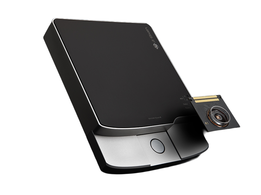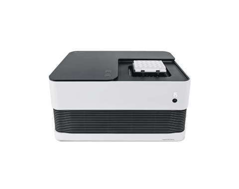
Our microchips, whether inside CorePlate™ single-well or integrated in CorePlate™ multi-wells, gather high-resolution electrophysiological imaging data from large networks of cultured neurons. Experimenters can browse the activity of spontaneous and chemically induced spatio-temporal patterns, watch the neural network’s response to stimuli and even study complex network topology.
Here you can view a selection of MEA recordings made with 3Brain neural recording systems:
200 ms of spontaneous electrophysiological activity of hippocampal cell cultures (22 days in vitro) showing sustained burst propagations over the whole network.
100 ms of spontaneous electrophysiological activity of hippocampal cell cultures (15 days in vitro) seeded at a low cellular density in order to resolve cellular activity (~4,000 neurons).
Recording of electrically evoked responses (100 ms) of a cultured hippocampal network (days in vitro) using two (A, B) different on-chip stimulating electrodes. Even responses below 3 ms can be recorded without electrically induced artifacts.
Spiking activity recorded from primary cortical neuronal culture at DIV 25 with a 3D scaffolding.
Two different examples (A and B) of different activity patterns recorded from the same hippocampal culture (22 DIV) from a 2-day old post-natal rat.
Human iPSC-derived neuronal cultures show an increased firing activity and network synchronicity following maturation at different Days in Vitro.
Sustained, tonic electrical activity of a h-iPSCs cortical spheroid on a single-well HD-MEA at DIV 49.
Analysis of the trajectory propagation of the heartbeat from a h-IPSC derived cardiac spheroid plated on the HD-MEA after 104 days.
One second recording of the uncorrelated spontaneous spiking activity of multiple neuronal spheroids seeded on a single-well HD-MEA after 3 days.
Field potentials and multi-unit activities can be sensed with unprecedented spatio temporal detail with both our HD-MEA (microelectrode array) and CorePlate™ powered 6-well devices .The large field of view enables assays on large tissue portions, such as rat cortico-hippocampal slices or brain organoids, to be carried out.
LFP oscillatory events across an olfactory bulb slice of a mouse at 8 weeks old elicited by Mk-801 perfusion. The duration of the event is 500ms.
Recording of hippocampal activation from two different mice, respectively 21 (A) and 27 (B) post-natal days, under perfusion with 4-Amino-Pyridine.
Recording of a rat hippocampal slice (14 days old) during spontaneous activity (A) and after perfusion with 4-Aminopyridine (B immediately after; C: 15 min after perfusion).
Functional imaging of distinct inter-ictal (I-IC) events obtained by superimposing the neuronal activity, here recorded from a cortico-hippocampal slice on a 3Brain high-resolution microchip, with a micrograph of the slice itself.
Cerebellar slice recording from a mouse (21 days old), showing spontaneous electrophysiological activity in Purkinje cells.
Our microchips integrated in high-density microelectrode arrays (HD-MEAs) and in CorePlates™ multi-wells are ideal for investigating explanted retinas, thanks to their large-scale sensing areas (up to 5.1 x 5.1 mm²) that feature a temporal resolution which outperforms traditional imaging techniques.
Activity footprints of a ganglion cell (top) and a polyaxonal amacrine cell (bottom) from an ex-vivo mouse retina showing single (ganglion cell) and multiple (amacrine cell) axonal propagations.
Recording of light-elicited activity of salamander retinal ganglion cells.
Retinal ganglion cell axonal responses to flickering (1Hz) checkerboard stimulus under dark mesopic conditions. The recording clearly shows propagating impulses along axonal bundles (P113 days mouse).
Light-elicited ganglion cell responses of a mouse retina (P113) showing the activity of retinal ON ganglion cells after onset of a full field light stimulus (2.8 cd*s/m2). Immediately after stimulus onset, ON transient ganglion cell types respond to the light followed by more ON sustained ganglion cell types. The optic disc is in the middle, the dorsal side at the bottom and the ventral side at the top.
Spontaneous waves recorded from the ganglion cell layer in a P11 mouse retina. The activity becomes stronger in the presence of the GABA-A receptor antagonist bicuculline (10 µM) and furosemide (100 µM), a blocker of the potassium-chloride co-transporter KCC2.
Recording of spontaneous electrophysiological activity from the ganglion cell layer in the Cone Rod Homeobox knockout mouse retina, a model of photoreceptor dystrophy. Dystrophic retinas are characterized by pathological, strong spontaneous bursting and oscillations in the ganglion cell layer. In this example, bursts are generated in cell bodies and propagate along axons converging toward the optic disc.