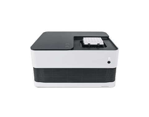
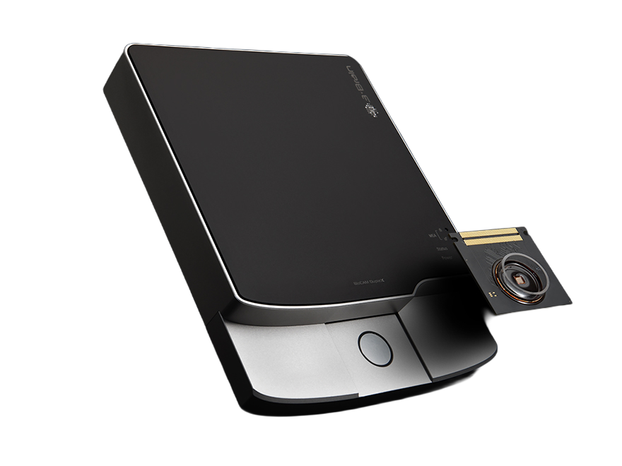
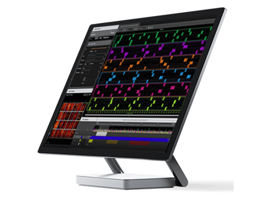
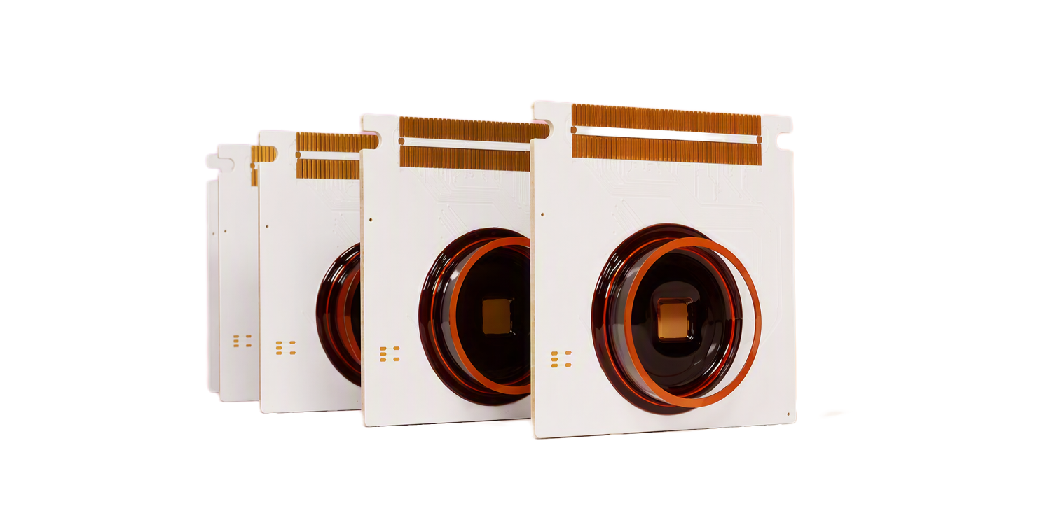
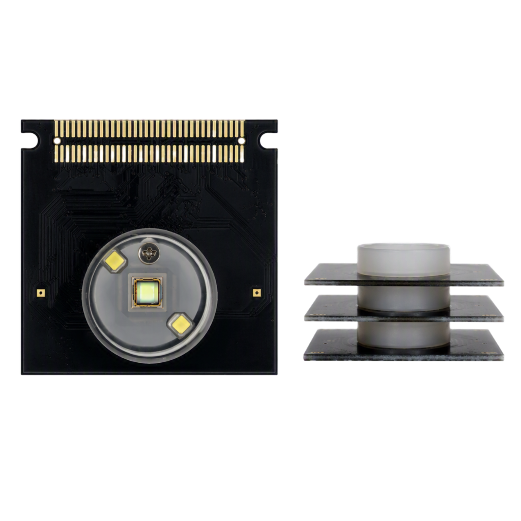
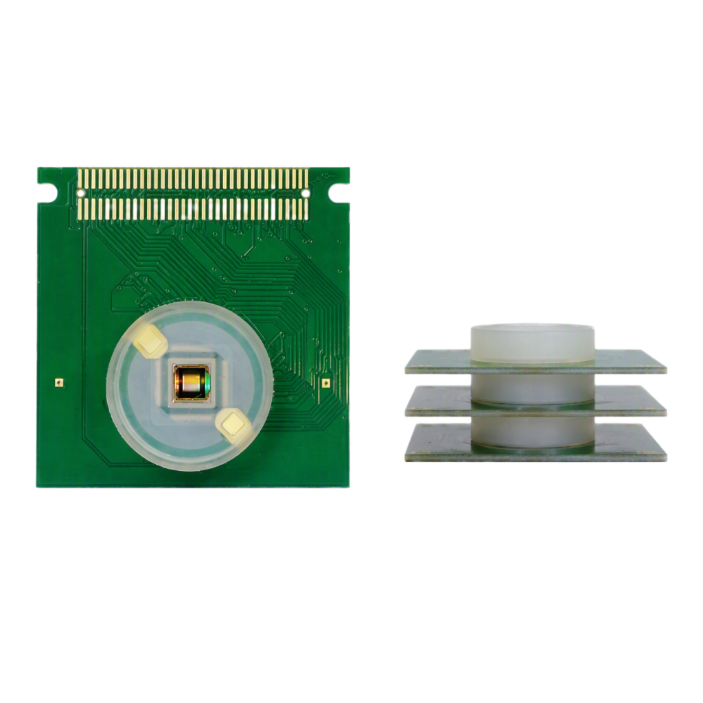
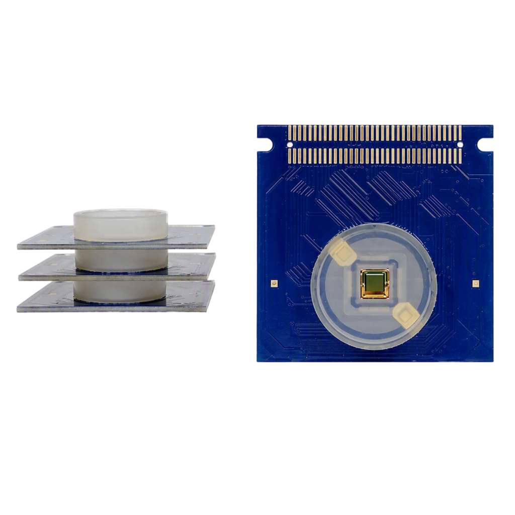
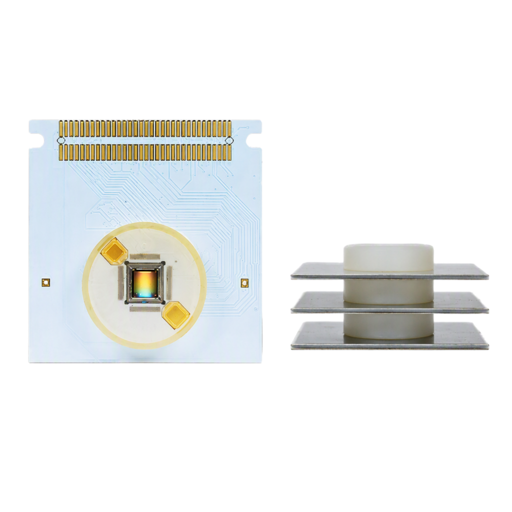
Our high-density microelectrode array (HD-MEA), also known as multielectrode array, features 4 096 low-noise microelectrodes that allow for simultaneous electrophysiological recording.

Investigating cellular networks requires time, expertise and effort to obtain the performance necessary to validate research hypotheses. With 3Brain as your technology partner, you can easily capture every cell signal in high resolution.
Our powerful HD-MEA (microelectrode array / multielectrode array) system collects high-density electrophysiological data from preparations made of brain slices, neuronal tissue, human-derived stem cells, brain organoids, retinal tissue and cardiac cultures and tissues.
Different HD-MEA chip models for our microelectrode arrays are available to match your experimental needs in terms of resolution, field of view and stimulation capabilities.
Electrodes are coated with platinum to provide a stable and fully biocompatible surface. The quality of the recording is ensured by the outstanding signal-to-noise ratio, as you can see from our CorePlate™ Example Recordings.


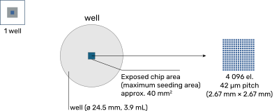
CorePlate™ 1W 27/42 is a high-density microelectrode array (HD-MEA) integrating 4 096 recording electrodes on a small and compact area of 2.67 mm × 2.67 mm, centered on a maximum seeding area of approx. 40 mm2. This arrangement is ideal in reducing the number of seeded cells.
Each microelectrode is 21 µm × 21 µm, with a pitch of 42 µm. The internal diameter of the reservoir is 24.5 mm, with a height of 9.1 mm.
The CorePlate™ 1W 27/42 is compatible with the BioCAM X and BioCAM DupleX.
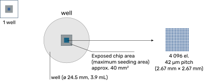
CorePlate™ 1W 27/42L has the same microelectrode recording array as the CorePlate™ 1W 27/42 (4 096 electrodes in a 2.67 mm × 2.67 mm area), but offers a larger surrounding exposed chip area (maximum seeding area) 40 mm2, which is ideal for positioning large tissues while guaranteeing optimal coupling with the microelectrode array.
Each microelectrode is 21 µm × 21 µm, with a pitch of 42 µm. The internal diameter of the reservoir is 24.5 mm, with a height of 9.1 mm.
The CorePlate™ 1W 27/42L is compatible with the BioCAM X and BioCAM DupleX.


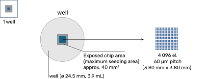
The CorePlate™1W 38/60 microelectrode array features 4 096 recording electrodes arranged in a 3.80 mm × 3.80 mm area, a perfect compromise for both cultures and brain tissues. Compared to previous versions, CorePlate™1W 38/60 achieves an improved signal-to-noise ratio by offering a recording mode where a subset electrode area can be accessed for ultra-low noise performance.
Moreover, every single recording electrode is bidirectional and can be used to release an electrical signal to your sample. The CorePlate™1W 38/60 provides a 3.80 mm × 3.8 mm grid of 4 096 electrodes for spatially accurate electrical stimulation at a cellular resolution.
Each microelectrode is 21 µm × 21 µm, with a 60 µm pitch. The internal diameter of the reservoir is 24.5 mm, with a height of 9.1 mm.
The CorePlate™1W 38/60 is compatible with the BioCAM DupleX only.
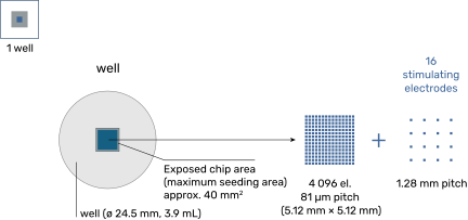
The CorePlate™ 1W 51/81 features a large sensing area (5.12 mm × 5.12 mm) with an increased electrode pitch of 81 µm. This makes it possible to record electrophysiological activity from large portions of neuronal tissue while maintaining a high spatial resolution.
The CorePlate™ 1W 51/81 also features a secondary array for stimulation with 16 microelectrodes, arranged in a 4 × 4 uniform grid, that are interspersed between the recording electrodes.
The electrode size for both recording and stimulating electrodes is 21 µm × 21 µm. The internal diameter of the reservoir is 24.5 mm, with a height of 9.1 mm.
CorePlate™ 1W 51/81 is compatible with both the BioCAM X and BioCAM DupleX. CorePlate™ 1W 51/81 supports seamless recording with the BioCAM DupleX. When paired with the BioCAM X, both recording and stimulation are fully enabled


Mode of operation
Recording
Number of electrodes
4 096, 64 × 64 grid
Number of simultaneously recorded electrodes
4 096
-
Electrode size
21 µm × 21 µm
Electrode pitch
42 µm
Electrode array dimensions (sensed area)
2.67 mm × 2.67 mm
Electrode Material
Pt black
Physical Specifications
Exposed chip area (maximum seeding area)
~ 40 mm2
Maximum well volume
3.9 mL
Well internal depth
9.1 mm
Well internal opening dimension
Ø 24.5 mm
Plate dimensions (W × D × H)
54.0 mm × 54.0 mm × 10.7 mm
(2.13 in × 2.13 in × 0.42 in)
Mode of operation
Recording and stimulation (bidirectional)
Number of electrodes
4 096, 64 × 64 grid
Number of simultaneously recorded electrodes
4 096
Number of stimulating electrodes
4 096
Electrode size
21 µm × 21 µm
Electrode pitch
60 µm
Electrode array dimensions (sensed area)
3.80 mm × 3.80 mm
Electrode Material
Pt black
Physical Specifications
Exposed chip area (maximum seeding area)
~ 40 mm2
Maximum well volume
3.9 mL
Well internal depth
9.1 mm
Well internal opening dimension
Ø 24.5 mm
Plate dimensions (W × D × H)
54.0 mm × 54.0 mm × 10.7 mm
(2.13 in × 2.13 in × 0.42 in)
Mode of operation
Recording
Number of electrodes
4 096, 64 × 64 grid
Number of simultaneously recorded electrodes
4 096
Number of stimulating electrodes
-
Electrode size
21 µm × 21 µm
Electrode pitch
81 µm
Electrode array dimensions (sensed area)
5.12 mm × 5.12 mm
Pt black
Dedicated Stimulation Array
Mode of operation
Stimulation
Number of electrodes
16 arranged in a 4 × 4 grid and uniformly interspersed between main array
Electrode size
21 µm × 21 µm
Electrode pitch
1.28 mm
Electrode array dimensions
3.86 mm × 3.86 mm
Physical Specifications
Exposed chip area (maximum seeding area)
~ 40 mm2
Maximum well volume
3.9 mL
Well internal depth
9.1 mm
Well internal opening dimension
Ø 24.5 mm
Plate dimensions (W × D × H)
54.0 mm × 54.0 mm × 10.7 mm
(2.13 in × 2.13 in × 0.42 in)