


Acute Retina
(paper)
Saltatory axonal conduction in the avian retina
J. Physiol. (2025). DOI: /10.1101/2022.07.04.498722

2025
Keywords:
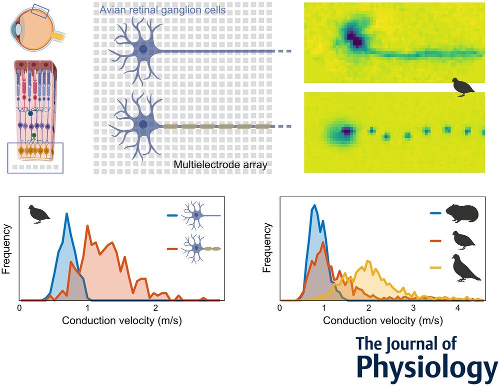
In contrast to most parts of the vertebrate nervous system, ganglion cell axons in the retina typically lack myelination. In the majority of species, ganglion cell axons only become myelinated after leaving the retina to form the optic nerve. The avian retina, however, presents a remarkable exception in that ganglion cell axons are partly myelinated in the retinal nerve fibre layer. It was hypothesized that the optically detrimental properties of retinal myelination are evolutionarily offset by advantages in spike conduction velocity. Using high-resolution multielectrode array recordings, we analysed the spike conduction in the retina of various avian species in comparison to mammalian species. Indeed, mammals showed lower conduction velocities than avian species. Myelinated axons typically achieved higher conduction velocities than unmyelinated axons. Notably, some myelinated axons exhibited conduction velocities lower than those of unmyelinated axons. Anatomical analyses revealed that myelination in the nerve fibre layer was accompanied by the formation of nodes of Ranvier. The internode length was positively correlated with the axon diameter. In physiological recordings, the spatial extent of simultaneously active nodes was positively correlated with the conduction velocity. Conversely, the internode length and the activation kinetics of a node were weak predictors of conduction velocity. Overall, this study illuminates the unique features of the avian retina and offers insights into the functional requirements and evolutionary pressures of myelination affecting conduction velocity in the nervous system.
Key points
- Intraretinal saltatory axonal spike conduction was studied across multiple avian species using high-resolution multielectrode arrays.
- The highest conduction velocities were observed exclusively in saltatory axons, while the lowest were found in non-saltatory axons. However, slow myelinated axons exist, exhibiting a surprisingly large overlap in conduction velocities with unmyelinated axons.
- The spatial extent of a spike showed a strong positive correlation with conduction velocity.
- The internodal length exhibited a positive correlation with axon diameter, and the variability in internodal length was smaller within individual axons than across axons.
- Intraretinal axonal conduction velocities across species appear to align with their ecological niche. The maximal intraretinal spike conduction velocity observed in birds was up to four times faster than in rodents.

Brain Slices
(paper)
Unsupervised pipeline for the identification of cortical excitatory and inhibitory neurons in high-density multielectrode arrays with ground-truth validation
eLife (2025). DOI: 10.7554/eLife.106557.3
2025
Keywords:
Large-scale extracellular recording techniques have advanced the study of neuronal circuits but lack methods to reliably identify cell types while scaling to thousands of neurons. We introduce spikeMAP, a pipeline for analyzing large-scale in vitro cortical recordings that combines spike sorting with cell-type identification using viral and optogenetic validation. SpikeMAP integrates data analysis with optogenetic, viral, and pharmacological protocols to dynamically probe distinct cell types while recording from large populations. The pipeline fits spike waveforms using spline interpolation to measure half-amplitude and peak-to-peak durations, applies principal component analysis and k-means clustering to isolate single-neuron signals, and uses linear discriminant analysis to optimize cluster separability. Channel source locations are determined through spatiotemporal spike waveform characteristics. Applied to mouse prefrontal cortex slices recorded on a 4096-channel array, spikeMAP effectively distinguishes regular-spiking excitatory neurons from fast-spiking inhibitory interneurons via action potential waveform, Fano factor, and spatial cross-correlations. This validated toolbox enables comprehensive characterization of neuronal activity across cell types in high-density recordings, offering a scalable approach to study microcircuit interactions in the brain.

Brain Slices
(paper)
Transplanted human striatal progenitors exhibit functional integration and modulate host circuitry in a Huntington’s disease animal model
Pharmacological Research (2025). DOI: 10.1016/j.phrs.2025.107905.
2025
Keywords:
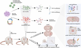
Huntington’s disease (HD) is a fatal neurodegenerative disorder caused by a CAG repeat expansion in the HTT gene. This leads to progressive loss of striatal neurons and motor-cognitive decline. While current gene-targeting approaches aiming at reducing somatic instability show promise – especially in case of early treatment – they cannot restore the already compromised neuronal circuitry at advanced disease stages. Thus, cell replacement therapy offers a regenerative strategy to rebuild damaged striatal circuits. Here, we report that human striatal progenitors (hSPs) derived from embryonic stem cells via a morphogen-guided protocol survive long-term when transplanted into a rodent model of HD and recapitulate key aspects of ventral telencephalic development. By employing single-nucleus RNAseq of the grafted cells, we resolved their transcriptional profile with unprecedented resolution. This has identified transcriptional signals of D1- and D2-type medium spiny neurons (MSN), Medial Ganglionic Eminence (MGE) and Caudal Ganglionic Eminence (CGE) -derived interneurons, and regionally specified astrocytes. Moreover, we demonstrate that grafted cells undergo further maturation 6 months post-transplantation, acquiring the expected regionally defined transcriptional identity. Immunohistochemistry confirmed stable graft composition over time and supported a neurogenic-to-gliogenic switch post-transplantation. Multiple complementary techniques including virus-based tracing and electrophysiology assays demonstrated anatomical and functional integration of the grafts. Notably, chemogenetic modulation of graft activity regulated striatal-dependent behaviors, further supporting effective graft integration into host basal ganglia circuits. Altogether, these results provide preclinical evidence that hSP-grafts can reconstruct striatal circuits and modulate functionally relevant behaviors. The ability to generate a scalable, molecularly defined progenitor population capable of in vivo functional integration supports the potential of hSPs for clinical application in HD and related basal ganglia disorders.

Brain Slices
(paper)
Dynamic mapping of network-level LTP in the hippocampus via high-resolution bioelectrical sensing
APL Bioeng. (2025). DOI: /10.1063/5.0258985
2025
Keywords:
Understanding the complexity of neural network dynamics demands advanced biosensing technologies capable of capturing large-scale interactions with high spatial and temporal precision. Traditional approaches, such as patch-clamp and field recordings, are inherently limited in resolving network-wide synaptic connections, particularly long-term potentiation (LTP), due to their localized scope and indirect access to hippocampal subfields. To address these challenges, we introduce EvoNES, a CMOS-based high-definition 4096 microelectrode array platform that leverages bidirectional stimulus-responsive biosensing functionality. By coupling precise external electrode stimulation targeting the Schaffer collateral and medial perforant pathways with simultaneous on-chip bioelectrical recordings, EvoNES enables the first real-time quantification of evoked responses and LTP dynamics across the entire hippocampal circuit. This system bridges critical gaps in traditional techniques, providing a mesoscopic-scale view of cell assemblies interplay and delivering unprecedented insights into the distributed mechanisms underlying memory encoding and learning processes. Advanced computational analyses generate variation maps revealing distinct voltage fluctuation patterns and differential sensitivity across hippocampal subregions during synaptic potentiation. Our findings identify four distinct waveform classes within the CA1–CA3 network and three unique evoked firing patterns in the dentate gyrus (DG). Post-tetanic responses show faster induction, expanded activated zones, and the activation of previously silent cell assemblies, indicating significant network restructuring. Applied in aged mice, EvoNES demonstrates age-dependent changes in network LTP, both quantitatively and qualitatively. This high-resolution biosensing platform in a live neural context provides unprecedented insights into hippocampal memory formation and offers a powerful tool for investigating neural plasticity and network interactions in both health and disease states.

Neuronal Cultures
(paper)
The lncRNA Gas5 is an activity-responsive scaffold that mediates cAMP-dependent synaptic plasticity
Sci. Signal. (2025). DOI: 10.1126/scisignal.adn2044
2025
Keywords:
Changes in the transcriptome are critical in shaping the structural plasticity of neurons, which underpins learning and long-term memory storage. Here, we explored the effect of two opposing, plasticity-associated pathways—cAMP second-messenger signaling and metabotropic glutamate receptor (mGluR1 and mGluR5) signaling—on the transcriptome in hippocampal neurons and how these pathways operate in distinct and coordinated manners to induce structural changes. Integration of transcriptome data and molecular pathway analysis identified central “hub” genes that were rapidly induced by cAMP and/or mGluR1/5 in hippocampal neurons. These included the long noncoding RNA (lncRNA) Gas5, whose expression was induced specifically by cAMP and which was targeted to dendrites by the kinesin motor protein KIF1A. In the dendrites, Gas5 interacted with various proteins and coding and noncoding RNAs associated with synaptic function and plasticity, and these interactions were altered by cAMP signaling. Gas5 interacted with the microRNA miR-26a-5p and sequestered it from several of its mRNA targets associated with neuronal function and whose translation was induced by cAMP. Gas5 was critical for excitatory synaptic transmission induced by cAMP but not those induced by mGluR1/5. Furthermore, Gas5 deficiency impaired dendritic branching and synapse morphology, and Gas5 abundance was decreased in the hippocampus of a mouse model of Alzheimer’s disease. Together, these findings provide insight into the transcriptional networks involved in synaptic plasticity and a lncRNA interactome that mediates dendritically localized regulation of excitatory synaptic transmission and neuronal architecture.

Neuronal Cultures
(paper)
CACNA1A loss-of-function affects neurogenesis in human iPSC-derived neural models
Cell. Mol. Life Sci. (2025). DOI: 10.1007/s00018-025-05740-7
2025
Keywords:
CACNA1A encodes the pore-forming α1A subunit of the CaV2.1 calcium channel, whose altered function is associated with various neurological disorders, including forms of ataxia, epilepsy, and migraine. In this study, we generated isogenic iPSC-derived neural cultures carrying CACNA1A loss-of-function mutations differently affecting CaV2.1 splice isoforms. Morphological, molecular, and functional analyses revealed an essential role of CACNA1A in neurodevelopmental processes. We found that different CACNA1A loss-of-function mutations produce distinct neurodevelopmental deficits. The F1491S mutation, which is located in a constitutive domain of the channel and therefore causes a complete loss-of-function, impaired neural induction at very early stages, as demonstrated by changes in single-cell transcriptomic signatures of neural progenitors, and by defective polarization of neurons. By contrast, cells carrying the Y1854X mutation, which selectively impacts the synaptically-expressed CaV2.1[EFa] isoform, behaved normally in terms of neural induction but showed altered neuronal network composition and lack of synchronized activity. Our findings reveal previously unrecognized roles of CACNA1A in the mechanisms underlying neural induction and neural network dynamics and highlight the differential contribution of the divergent variants CaV2.1[EFa] and CaV2.1[EFb] in the development of human neuronal cells.

Brain Slices
(paper)
Neurodevelopmental origins of structural and psychomotor defects in CXCR4-linked primary immunodeficiency
Neuron (2025). DOI: 10.1016/j.neuron.2025.05.016
2025
Keywords:
Inborn errors of immunity (IEI), as congenital chronic disorders, are often associated with neurobehavioral symptoms, traditionally considered secondary to patient burden. Their origin, however, has yet to be addressed. Here, we found that IEI-associated genes are expressed in neural lineages during human brain development, and in the absence of immunological challenges, IEI mutations directly impair neurodevelopmental trajectories, leading to psychomotor defects. Warts hypogammaglobulinemia immunodeficiency myelokathexis (WHIM) mice—bearing a mutation causing Cxcr4 hyperactivation—show developmental foliation defects of the cerebellum correlating with sensorimotor and affective dysfunctions, which recapitulate the alterations described in patients. WHIM cerebella single-cell profiling revealed major transcriptional deregulation in granule cell progenitors, whose aberrant proliferation and migration induce foliation and circuit defects. AMD3100 intracerebroventricular injection rescues both morphological and behavioral defects, demonstrating their brain-specific and Cxcr4-dependent origin. Collectively, our findings highlight the relevance of neurodevelopmental implications underlying psychomotor IEI manifestations, broadening our understanding of these conditions beyond immune dysfunctions.

Acute Retina
(paper)
Filter-based models of suppression in retinal ganglion cells: Comparison and generalization across species and stimuli
PLoS Comput Biol (2025). DOI: /10.1371/journal.pcbi.1013031
2025
Keywords:
The dichotomy of excitation and suppression is one of the canonical mechanisms explaining the complexity of neural activity. Computational models of the interplay of excitation and suppression in single neurons aim at investigating how this interaction affects a neuron’s spiking responses and shapes the encoding of sensory stimuli. Here, we compare the performance of three filter-based stimulus-encoding models for predicting retinal ganglion cell responses recorded from axolotl, mouse, and marmoset retina to different types of temporally varying visual stimuli. Suppression in these models is implemented via subtractive or divisive interactions of stimulus filters or by a response-driven feedback module. For the majority of ganglion cells, the subtractive and divisive models perform similarly and outperform the feedback model as well as a linear-nonlinear (LN) model with no suppression. Comparison between the subtractive and the divisive model depends on cell type, species, and stimulus components, with the divisive model generalizing best across temporal stimulus frequencies and visual contrast and the subtractive model capturing in particular responses for slow temporal stimulus dynamics and for slow axolotl cells. Overall, we conclude that the divisive and subtractive models are well suited for capturing interactions of excitation and suppression in ganglion cells and perform best for different temporal regimes of these interactions.

Brain Slices
(paper)
MEA-seqX: High-Resolution Profiling of Large-Scale Electrophysiological and Transcriptional Network Dynamics
Adv. Sci. (2025). DOI: 10.1002/advs.202412373
2025
Keywords:
Concepts of brain function imply congruence and mutual causal influence between molecular events and neuronal activity. Decoding entangled information from concurrent molecular and electrophysiological network events demands innovative methodology bridging scales and modalities. The MEA-seqX platform, integrating high-density microelectrode arrays, spatial transcriptomics, optical imaging, and advanced computational strategies, enables the simultaneous recording and analysis of molecular and electrical network activities at mesoscale spatial resolution. Applied to a mouse hippocampal model of experience-dependent plasticity, MEA-seqX unveils massively enhanced nested dynamics between transcription and function. Graph–theoretic analysis reveals an increase in densely connected bimodal hubs, marking the first observation of coordinated hippocampal circuitry dynamics at molecular and functional levels. This platform also identifies different cell types based on their distinct bimodal profiles. Machine-learning algorithms accurately predict network-wide electrophysiological activity features from spatial gene expression, demonstrating a previously inaccessible convergence across modalities, time, and scales.

Brain Slices
(paper)
A novel prefrontal cortex and hippocampus combined brain slice based on in vivo diffusion tensor imaging of healthy male rats
Neurosci. Lett. (2025). DOI: 10.1016/j.neulet.2025.138171.
2025
Keywords:
The pathway between the prefrontal cortex (PFC) and hippocampus (HPC) has been associated with various psychiatric disorders. While hippocampal brain slices are extensively utilized, their use has traditionally been constrained in studying long connectivity between PFC and HPC due to nerve fiber rupture during the slicing process. Consequently, optimizing brain slice preparation is crucial. The experiment consisted of three phases. Initially, the structural connection of the PFC-HPC pathway was examined using diffusion tensor imaging (DTI) data from healthy male rats. Subsequently, combined PFC-HPC brain slices were created through vibratome based on imaging acquisition. Finally, the morphology and electrophysiology of the combined brain slices were analyzed. DTI findings revealed numerous nerve fibers linking the two brain regions in the rat brain. Subsequently, a successful preparation of combined PFC-HPC brain slices cut at a 7 – 8° angle relative to the middle sagittal plane was achieved using a vibratome. Hematoxylin and eosin staining results confirmed that PFC-HPC fibers remained well-preserved in the combined brain slice. Electrophysiological recordings indicated that synchronized neuronal activity occurred in the HPC upon PFC stimulation, which depended on hippocampal activity and the integrity of PFC-to-HPC connectivity. A novel procedure for the successful preparation of healthy combined HPC-PFC brain slices, maintaining a complete fiber bundle connection between PFC and HPC, is proposed. This methodology enhances the understanding of the preservation of PFC-HPC connectivity in specific angled brain slice preparations, thereby facilitating neuroscience research focused on the longrange circuitry of subregions of interest.

Neuronal Cultures
Criticality in neural cultures: Insights into memory and connectivity in entorhinal-hippocampal networks
Chaos Solit. Fractals (2025). DOI: 10.1016/j.chaos.2025.116184
2025
Keywords:
The brain is a complex system of interconnected regions that underlie memory, cognition, and perception. Today, our understanding of the brain’s dynamic processes remains incomplete, particularly regarding differences in electrophysiological activity and inter-regional connectivity among specific areas. To explore this, we investigated the electrical activity, functional connectivity, and interactions of neural cultures differentiated into hippocampal, isocortical, and entorhinal networks using multi-electrode arrays (MEAs) to record extracellular local field potentials. Our results showed that collective synchronization events, or network bursts, were present in all cultures except for the hippocampal networks. Interestingly, introducing entorhinal neuron spheroids onto hippocampal cultures induced synchronized activity. Furthermore, Self-organized criticality analysis confirmed that all networks, except hippocampal cultures, were in a critical regime. Moreover, we found that entorhinal-hippocampal coupling facilitated criticality, promoting recurrent synchronized activity patterns. The consistent scaling exponents across configurations underscore the universality of criticality in biological networks. Finally, power spectrum analysis revealed a theta band peak in connected entorhinal-hippocampal cultures, consistent with in vivo studies, highlighting the role of theta oscillations in memory consolidation. Our findings provide more insights into brain functioning and offer an in vitro model for studying learning and memory.

Acute Retina
(paper)
Spatial distribution and functional integration of displaced retinal ganglion cells
Sci. Rep. (2025). DOI: 10.1038/s41598-025-91045-5
2025
Keywords:
The retina contains distinct types of ganglion cells, which form mosaics with cells of each type at each position of the visual field. Displaced retinal ganglion cells (dRGCs) occur with cell bodies in the inner nuclear layer (INL), and regularly placed RGCs with cell bodies in the ganglion cell layer. An example of mammalian dRGCs are M1-type intrinsically photosensitive ganglion cells (ipRGCs). Little is known, however, about their relationship with regularly placed ipRGCs. We identified mouse ipRGC types M1, M2, and M4/sONɑ by immunohistochemistry and light microscopy. Reconstruction of immunolabeled mosaics from M1 and sONɑ RGCs indicated that dRGCs tiled the retina with their regular RGC partners. Multi-electrode array recordings revealed conventional receptive fields of displaced sONɑ RGCs which fit into the mosaic of their regular counterparts. An RGC distribution analysis showed type-specific dRGC patterns which followed neither the global density distribution of all RGCs nor the local densities of corresponding cell types. The displacement of RGC bodies into the INL occurs in a type-dependent manner, where dRGCs are positioned to form complete mosaics with their regular partners. Our data suggest that dRGCs and regular RGCs serve the same functional role within their corresponding population of RGCs.
Acute Retina
(paper)
Homologous amacrine to amacrine gap junction coupling serves communication between neighbour OFF alpha retinal ganglion cells
J. Physiol. (2025). DOI: 10.1113/JP287699
2025
Keywords:

Multiplexed visual coding by retinal ganglion cells (RGCs) has gained much support. Mouse transient OFF alpha RGCs (tOFFα RGCs) are excellent subjects to study this issue as they form direct RGC–RGC gap junctions (GJs) that serve spike synchronization, population coding and likely information multiplexing. In addition, tOFFα RGCs maintain GJs with a population of wide-field amacrine cells (ACs) that have been suspected to mediate an additional, loose medium-scale correlation of tOFFα RGC spikes. However, the spatial and temporal constraints of the GJ-mediated AC–RGC signalling have yet to be tested directly via a combination of morphological and functional approaches. Here we show that AC-mediated medium-scale spike correlations are strongly related to spike bursts. On the other hand, our data also show that coupled ACs’ somata form spatially separated clusters each overlapping with only a single tOFFα RGC dendritic arbour suggesting the existence of GJ-coupled tOFFα RGC–AC functional units. This finding seemingly argues against the hypothesis that ACs distribute common noise for burst-based medium-scale RGC spike correlations. However, we also found a high incidence of AC–AC GJ connections thereby forming the morphological substrate for the interconnection of functional units to correlate spike bursts on a medium time scale. These data thus suggest that besides encoding visual information by a single cell, tOFFα RGCs utilize RGC–RGC GJs to directly connect RGCs as well as AC–AC GJs to interconnect tOFFα RGC functional units to mediate two forms of population codes via precise spike synchronization and loose burst correlations, respectively.

Acute Retina
(paper)
Pivotal roles of melanopsin containing retinal ganglion cells in pupillary light reflex in photopic conditions
Front. Cell. Neurosci. (2025). DOI: 10.3389/fncel.2025.1547066
2025
Keywords:
The pupillary light reflex (PLR) is crucial for protecting the retina from excess light. The intrinsically photosensitive retinal ganglion cells (ipRGCs) in the retina are neurons that are critical to generating the PLR, receiving rod/cone photoreceptor signals and directly sensing light through melanopsin. Previous studies have investigated the roles of photoreceptors and ipRGCs in PLR using genetically-modified mouse models. Herein, we acutely ablated photoreceptors using N-nitroso-N-methylurea (MNU) to examine the roles of ipRGCs in the PLR. We conducted PLR and multiple electrode array (MEA) recordings evoked by three levels of light stimuli before and 5 days after MNU intraperitoneal (i.p..) injection using C57BL6/J wildtype (WT) mice. We also conducted these measurements using the rod & cone dysfunctional mice (Gnat1–/–& Cnga3–/–:dKO) to compare the results to published studies in which mutant mice were used to show the role of photoreceptors and ipRGCs in PLR. PLR pupil constriction increased as the light stimulus intensified in WT mice. In MNU mice, PLR was not induced by the low light stimulus, suggesting that photoreceptors induced the PLR at this light intensity. By contrast, the high light stimulus fully induced PLR, similar to the response in WT mice. In dKO mice, no PLR was evoked by the low-light stimulus and a slow-onset PLR was evoked by the high-light stimulus, consistent with previous reports. Ex vivo MEA recording in the MNU tissue revealed a population of ipRGCs with a fast onset and peak time, suggesting that they drove the fast PLR response. These results suggest that ipRGCs primarily contribute to the PLR at a high light intensity, which does not agree with the previous results shown by mutant mouse models. Our results indicate that the melanopsin response in ipRGCs generate fast and robust PLR when induced by high light.

Acute Retina
(paper)
A membrane-targeted photoswitch restores physiological ON/OFF responses to light in the degenerate retina
Nat. Commun. (2025). DOI: 10.1038/s41467-025-55882-2
2025
Keywords:
The lack of effective therapies for visual restoration in Retinitis pigmentosa and macular degeneration has led to the development of new strategies, such as optogenetics and retinal prostheses. However, visual restoration is poor due to the massive light-evoked activation of retinal neurons, regardless of the segregation of visual information in ON and OFF channels, which is essential for contrast sensitivity and spatial resolution. Here, we show that Ziapin2, a membrane photoswitch that modulates neuronal capacitance and excitability in a light-dependent manner, is capable of reinstating, in mouse and rat genetic models of photoreceptor degeneration, brisk and sluggish ON, OFF, and ON-OFF responses in retinal ganglion cells evoked by full-field stimuli, with reactivation of their excitatory and inhibitory conductances. Intravitreally injected Ziapin2 in fully blind rd10 mice restores light-driven behavior and optomotor reflexes. The results indicate that Ziapin2 is a promising molecule for reinstating physiological visual responses in the late stages of retinal degeneration.

Technology
(paper)
Longitudinal and Noninvasive Intracellular Recordings of Spontaneous Electrophysiological Activity in Rat Primary Neurons on Planar MEA Electrodes
Advanced Materials (2025). DOI: 10.1002/adma.202412697
2025
Keywords:
Presently, the in vitro recording of intracellular neuronal signals on microelectrode arrays (MEAs) requires complex 3D nanostructures or invasive and approaches such as electroporation. Here, it is shown that laser poration enables intracellular coupling on planar electrodes without damaging neurons or altering their spontaneous electrophysiological activity, allowing the process to be repeated multiple times on the same cells. This capability distinguishes laser-based neuron poration from more invasive methods like electroporation, which typically serve as endpoint measurement for cells. It is demonstrated that planar MEA electrodes, when combined with laser cell optoporation and live cell staining, can record spontaneous intracellular signaling from primary neurons in vitro. This approach allows for the detection of attenuated signals resembling positive monophasic intracellular action potentials. Recordings after laser optoporation also reveal subthreshold signals such as post-synaptic potentials that are essential for assessing neuronal network plasticity and connectivity. Moreover, the noninvasiveness of the process enables repeated intracellular recordings over multiple days from the same cells.

Neuronal Cultures
(paper)
Dual inhibition of MAPK/ERK and BMP signaling induces entorhinal-likeidentity in mouse ESC-derived pallial progenitors
Stem Cell Reports (2025). DOI: 10.1016/j.stemcr.2024.12.002
2025
Keywords:
.jpeg)
Highlights
- MAPK/ERK and BMP inhibition (MiBi) specifies an entorhinal-like identity
- MiBi neurons activate a distinct gene expression profile for neuronal connectivity
- MiBi and isocortical neurons show different connectivity with hippocampal cells
- MiBi/hippocampal functional assembloids develop spontaneous theta activity
Summary
The mechanisms that determine distinct embryonic pallial identities remain elusive. The central role of Wnt signaling in directing dorsal telencephalic progenitors to the isocortex or hippocampus has been elucidated. Here, we show that timely inhibition of MAPK/ERK and BMP signaling in neuralized mouse embryonic stem cells (ESCs) specifies a cell identity characteristic of the allocortex. Comparison of the global gene expression profiles of neural cells generated by MAPK/ERK and BMP inhibition (MiBi cells) with those of cells from early postnatal encephalic regions reveals a pallial identity of MiBi cells, distinct from isocortical and hippocampal cells. MiBi cells display a unique pattern of gene expression and connectivity, and share molecular and electrophysiological features with the entorhinal cortex. Our results suggest that early changes in cell signaling can specify distinct pallial fates that are maintained by specific neuronal lineages independent of subsequent embryonic morphogenetic interactions and can determine their functional connectivity.

Neuronal Cultures
(paper)
High-density multielectrode arrays bring cellular resolution to neuronal activity and network analyses of corticospinal motor neurons
Sci. Rep. (2025). DOI: 10.1038/s41598-024-83883-6
2025
Keywords:
Corticospinal motor neurons (CSMN), located in the motor cortex of the brain, are one of the key components of the motor neuron circuitry. They are in part responsible for the initiation and modulation of voluntary movement, and their degeneration is the hallmark for numerous diseases, such as amyotrophic lateral sclerosis (ALS), hereditary spastic paraplegia, and primary lateral sclerosis. Cortical hyperexcitation followed by in-excitability suggests the early involvement of cortical dysfunction in ALS pathology. However, a high-spatiotemporal resolution on our understanding of their functional health and connectivity is lacking. Here, we combine optical imaging with high-density microelectrode array (HD-MEA) system enabling single cell resolution and utilize UCHL1-eGFP mice to bring cell-type specificity to our understanding of the electrophysiological features of healthy CSMN, as they mature and form network connections with other cortical neurons, in vitro. This novel approach lays the foundation for future cell-type specific analyses of CSMN that are diseased due to different underlying causes with cellular precision, and it will allow the assessment of their functional response to compound treatment, especially for drug discovery efforts in upper motor neuron diseases.

Brain Slices
(paper)
Network-wide effects of pallidal deep brain stimulation normalised abnormal cerebellar cortical activity in the dystonic animal model
Neurobiol. Dis. (2025). DOI: 10.1016/j.nbd.2024.106779
2025
Keywords:
Background
Deep brain stimulation (DBS) targeting globus pallidus internus (GPi) is a recognised therapy for drug-refractory dystonia. However, the mechanisms underlying this effect are not fully understood. This study explores how pallidal DBS alters spatiotemporal pattern formation of neuronal dynamics within the cerebellar cortex in a dystonic animal model, the dtsz hamster.
Methods
We conducted in vitro analysis using a high-density microelectrode array (HD-MEA) in the cerebellar cortex. For investigating the spatiotemporal pattern, mean firing rates (MFR), interspike intervals (ISI), spike amplitudes, and cerebellar connectivity among healthy control hamsters, dystonic dtsz hamsters, DBS- and sham-DBS-treated dtsz hamsters were analysed. A nonlinear data-driven method characterised the low-dimensional representation of the patterns in MEA data.
Results
Our HD-MEA recordings revealed reduced MFR and spike amplitudes in the dtsz hamsters compared to healthy controls. Pallidal DBS induced network-wide effects, normalising MFR, spike amplitudes, and connectivity measures in hamsters, thereby countervailing these electrophysiological abnormalities. Additionally, network analysis showed neural activity patterns organised into communities, with higher connectivity in both healthy and DBS groups compared to dtsz.
Conclusions
These findings suggest that pallidal DBS exerts some of its therapeutic effects on dystonia by normalising neuronal activity within the cerebellar cortex. Our findings of reduced MFR and spike amplitudes in the dtsz hamsters could be a hint of a decrease in neuronal fibres and synaptic plasticity. Treatment with pallidal DBS led to cerebellar cortical activity similar to healthy controls, displaying the network-wide impact of local stimulation.

Brain Slices
(paper)
Disruption of the Autism-Associated Pcdh9 Gene Leads to Transcriptional Alterations, Synapse Overgrowth, and Defective Network Activity in the CA1
J. Neurosci. (2024). DOI: /10.1523/JNEUROSCI.0491-24.2024
2024
Keywords:
Protocadherins, a family of adhesion molecules with a crucial role in cell–cell interactions, have emerged as key players in neurodevelopmental and psychiatric disorders. In particular, growing evidence links genetic alterations in the protocadherin 9 (PCDH9) gene with autism spectrum disorder and major depressive disorder. Furthermore, Pcdh9 deletion induces neuronal defects in the mouse somatosensory cortex, accompanied by sensorimotor and memory impairment. However, the synaptic and molecular mechanisms of PCDH9 in the brain remain largely unknown, particularly concerning its impact on brain pathology. To address this question, we conducted a comprehensive investigation of PCDH9’s role in the male mouse hippocampus at the ultrastructural, biochemical, transcriptomic, electrophysiological, and network levels. We show that PCDH9 mainly localizes at glutamatergic synapses and its expression peaks in the first week after birth, a crucial time window for synaptogenesis. Strikingly, Pcdh9 KO neurons exhibit oversized presynaptic terminal and postsynaptic density in the CA1. Synapse overgrowth is sustained by the widespread upregulation of synaptic genes, as revealed by single-nucleus RNA-seq (snRNA-seq), and the dysregulation of key drivers of synapse morphogenesis, including the SHANK2/CORTACTIN pathway. At the functional level, these structural and transcriptional abnormalities result in increased excitatory postsynaptic currents (mEPSC) and reduced network activity in the CA1 of Pcdh9 KO mice. In conclusion, our work uncovers Pcdh9’s pivotal role in shaping the morphology and function of CA1 excitatory synapses, thereby modulating glutamatergic transmission within hippocampal circuits.
Neuronal Cultures
(paper)
The Alzheimer's Disease Risk Gene CD2AP Functions in Dendritic Spines by Remodeling F-Actin
J. Neurosci. (2024). DOI: 10.1523/JNEUROSCI.1734-23.2024
2024
Keywords:
CD2-associated protein (CD2AP) was identified as a genetic risk factor for late-onset Alzheimer's disease (LOAD). However, it is unclear how CD2AP contributes to LOAD synaptic dysfunction underlying AD memory deficits. We have shown that loss of CD2AP function increases β-amyloid (Aβ) endocytic production, but it is unknown whether it contributes to synapse dysfunction. As CD2AP is an actin-binding protein, it may also function in F-actin-rich dendritic spines, which are the excitatory postsynaptic compartments. Here, we demonstrate that CD2AP colocalizes with F-actin in dendritic spines of primary mouse cortical neurons of both sexes. Cell-autonomous depletion of CD2AP specifically reduces spine density and volume, resulting in a functional decrease in synapse formation and neuronal network activity. Postsynaptic reexpression of CD2AP, but not blocking Aβ production, is sufficient to rescue spine density. CD2AP overexpression increases spine density, volume, and synapse formation, while a rare LOAD CD2AP mutation induces aberrant F-actin spine-like protrusions without functional synapses. CD2AP controls postsynaptic actin turnover, with the LOAD mutation in CD2AP decreasing F-actin dynamicity. Our data support that CD2AP risk variants could contribute to LOAD synapse dysfunction by disrupting spine formation and growth by deregulating actin dynamics.

Brain Slices
(paper)
High-Quality Seizure-Like Activity from Acute Brain Slices Using a Complementary Metal-Oxide-Semiconductor High-Density Microelectrode Array System
J. Vis. Exp. (2024). DOI:10.3791/67065
2024
Keywords:
Complementary metal-oxide-semiconductor high-density microelectrode array (CMOS-HD-MEA) systems can record neurophysiological activity from cell cultures and ex vivo brain slices in unprecedented electrophysiological detail. CMOS-HD-MEAs were first optimized to record high-quality neuronal unit activity from cell cultures but have also been shown to produce quality data from acute retinal and cerebellar slices. Researchers have recently used CMOS-HD-MEAs to record local field potentials (LFPs) from acute, cortical rodent brain slices. One LFP of interest is seizure-like activity. While many users have produced brief, spontaneous epileptiform discharges using CMOS-HD-MEAs, few users reliably produce quality seizure-like activity. Many factors may contribute to this difficulty, including electrical noise, the inconsistent nature of producing seizure-like activity when using submerged recording chambers, and the limitation that 2D CMOS-MEA chips only record from the surface of the brain slice. The techniques detailed in this protocol should enable users to consistently induce and record high-quality seizure-like activity from acute brain slices with a CMOS-HD-MEA system. In addition, this protocol outlines the proper treatment of CMOS-HD-MEA chips, the management of solutions and brain slices during experimentation, and equipment maintenance.

Brain Slices
(paper)
DENOISING: Dynamic enhancement and noise overcoming in multimodal neural observations via high-density CMOS-based biosensors
Front. Bioeng. Biotechnol. (2024). DOI: /10.3389/fbioe.2024.1390108
2024
Keywords:
Large-scale multimodal neural recordings on high-density biosensing microelectrode arrays (HD-MEAs) offer unprecedented insights into the dynamic interactions and connectivity across various brain networks. However, the fidelity of these recordings is frequently compromised by pervasive noise, which obscures meaningful neural information and complicates data analysis. To address this challenge, we introduce DENOISING, a versatile data-derived computational engine engineered to adjust thresholds adaptively based on large-scale extracellular signal characteristics and noise levels. This facilitates the separation of signal and noise components without reliance on specific data transformations. Uniquely capable of handling a diverse array of noise types (electrical, mechanical, and environmental) and multidimensional neural signals, including stationary and non-stationary oscillatory local field potential (LFP) and spiking activity, DENOISING presents an adaptable solution applicable across different recording modalities and brain networks. Applying DENOISING to large-scale neural recordings from mice hippocampal and olfactory bulb networks yielded enhanced signal-to-noise ratio (SNR) of LFP and spike firing patterns compared to those computed from raw data. Comparative analysis with existing state-of-the-art denoising methods, employing SNR and root mean square noise (RMS), underscores DENOISING’s performance in improving data quality and reliability. Through experimental and computational approaches, we validate that DENOISING improves signal clarity and data interpretation by effectively mitigating independent noise in spatiotemporally structured multimodal datasets, thus unlocking new dimensions in understanding neural connectivity and functional dynamics.
Brain Slices
(paper)
A high-density multi-electrode platform examining the effects of radiation on in vitro cortical networks
Sci. Rep. (2024). DOI: 10.1038/s41598-024-71038-6
2024
Keywords:
Radiation therapy and stereotactic radiosurgery are common treatments for brain malignancies. However, the impact of radiation on underlying neuronal circuits is poorly understood. In the prefrontal cortex (PFC), neurons communicate via action potentials that control cognitive processes, thus it is important to understand the impact of radiation on these circuits. Here we present a novel protocol to investigate the effect of radiation on the activity and survival of PFC networks in vitro. Escalating doses of radiation were applied to PFC slices using a robotic radiosurgery platform at a standard dose rate of 10 Gy/min. High-density multielectrode array recordings of radiated slices were collected to capture extracellular activity across 4,096 channels. Radiated slices showed an increase in firing rate, functional connectivity, and complexity. Graph-theoretic measures of functional connectivity were altered following radiation. These results were compared to pharmacologically induced epileptic slices where neural complexity was markedly elevated, and functional connections were strong but remained spatially focused. Finally, propidium iodide staining revealed a dose-dependent effect of radiation on apoptosis. These findings provide a novel assay to investigate the impacts of clinically relevant doses of radiation on brain circuits and highlight the acute effects of escalating radiation doses on PFC neurons.

Organoids & Spheroids
(paper)
Generation of human region-specific brain organoids with medullary spinal trigeminal nuclei
Cell Stem Cell (2024). DOI: 10.1016/j.stem.2024.08.004
2024
Keywords:

Highlights
- Human medullary SpV-like organoids (hmSpVOs) are created from hPSCs
- hmSpVOs resemble the spinal trigeminal nucleus (SpV) of the dorsal medulla
- hmSpVOs exhibit structural and functional maturation in long-term culture
- Trigeminothalamic tracts are established between hmSpVOs and thalamic organoids
Summary
Brain organoids with nucleus-specific identities provide unique platforms for studying human brain development and diseases at a finer resolution. Despite its essential role in vital body functions, the medulla of the hindbrain has seen a lack of in vitro models, let alone models resembling specific medullary nuclei, including the crucial spinal trigeminal nucleus (SpV) that relays peripheral sensory signals to the thalamus. Here, we report a method to differentiate human pluripotent stem cells into region-specific brain organoids resembling the dorsal domain of the medullary hindbrain. Importantly, organoids specifically recapitulated the development of the SpV derived from the dorsal medulla. We also developed an organoid system to create the trigeminothalamic projections between the SpV and the thalamus by fusing these organoids, namely human medullary SpV-like organoids (hmSpVOs), with organoids representing the thalamus (hThOs). Our study provides a platform for understanding SpV development, nucleus-based circuit organization, and related disorders in the human brain.
.svg)


