


Brain Slices
(paper)
A deep generative adversarial network capturing complex spiral waves in disinhibited circuits of the cerebral cortex
BMC Neurosci. (2023) DOI: 10.1186/s12868-023-00792-6

2023
Keywords:
Background
In the cerebral cortex, disinhibited activity is characterized by propagating waves that spread across neural tissue. In this pathological state, a widely reported form of activity are spiral waves that travel in a circular pattern around a fixed spatial locus termed the center of mass. Spiral waves exhibit stereotypical activity and involve broad patterns of co-fluctuations, suggesting that they may be of lower complexity than healthy activity.
Results
To evaluate this hypothesis, we performed dense multi-electrode recordings of cortical networks where disinhibition was induced by perfusing a pro-epileptiform solution containing 4-Aminopyridine as well as increased potassium and decreased magnesium. Spiral waves were identified based on a spatially delimited center of mass and a broad distribution of instantaneous phases across electrodes. Individual waves were decomposed into “snapshots” that captured instantaneous neural activation across the entire network. The complexity of these snapshots was examined using a measure termed the participation ratio. Contrary to our expectations, an eigenspectrum analysis of these snapshots revealed a broad distribution of eigenvalues and an increase in complexity compared to baseline networks. A deep generative adversarial network was trained to generate novel exemplars of snapshots that closely captured cortical spiral waves. These synthetic waves replicated key features of experimental data including a tight center of mass, a broad eigenvalue distribution, spatially-dependent correlations, and a high complexity. By adjusting the input to the model, new samples were generated that deviated in systematic ways from the experimental data, thus allowing the exploration of a broad range of states from healthy to pathologically disinhibited neural networks.
Conclusions
Together, results show that the complexity of population activity serves as a marker along a continuum from healthy to disinhibited brain states. The proposed generative adversarial network opens avenues for replicating the dynamics of cortical seizures and accelerating the design of optimal neurostimulation aimed at suppressing pathological brain activity.

Brain Slices
(paper)
An in vitro model of drug-resistant seizures for selecting clinically effective antiseizure medications in Febrile Infection-Related Epilepsy Syndrome
Front. Neurol. (2023). DOI: 10.3389/fneur.2023.1129138
2023
Keywords:
Introduction
FIRES is a rare epileptic encephalopathy induced by acute unremitting seizures that occur suddenly in healthy children or young adults after a febrile illness in the preceding 2 weeks. This condition results in high mortality, neurological disability, and drug-resistant epilepsy. The development of new therapeutics is hampered by the lack of validated experimental models. Our goal was to address this unmet need by providing a simple tool for rapid throughput screening of new therapies that target pathological inflammatory mechanisms in FIRES. The model was not intended to mimic the etiopathogenesis of FIRES which is still unknown, but to reproduce salient features of its clinical presentation such as the age, the cytokine storm and the refractoriness of epileptic activity to antiseizure medications (ASMs).
Methods
We refined an in vitro model of mouse hippocampal/temporal cortex acute slices where drug-resistant epileptic activity is induced by zero Mg2+/100 μM 4-aminopirydine. Clinical evidence suggests that acute unremitting seizures in FIRES are promoted by neuroinflammation triggered in the brain by the preceding infection. We mimicked this inflammatory component by exposing slices for 30 min to 10 μg/ml lipopolysaccharide (LPS).
Results
LPS induced a sustained neuroinflammatory response, as shown by increased mRNA levels of IL-1β, CXCL1 (IL-8), TNF, and increased IL-1β/IL-1Ra ratio. Epileptiform activity was exacerbated by neuroinflammation, also displaying increased resistance to maximal therapeutic concentrations of midazolam (100 μM), phenytoin (50 μM), sodium valproate (800 μM), and phenobarbital (100 μM). Treatment of LPS-exposed slices with two immunomodulatory drugs, a mouse anti-IL-6 receptor antibody (100 μM) corresponding to tocilizumab in humans, or anakinra (1.3 μM) which blocks the IL-1 receptor type 1, delayed the onset of epileptiform events and strongly reduced the ASM-resistant epileptiform activity evoked by neuroinflammation. These drugs were shown to reduce ASM-refractory seizures in FIRES patients.
Discussion
The neuroinflammatory component and the pharmacological responsiveness of epileptiform events provide a proof-of-concept validation of this in vitro model for the rapid selection of new treatments for acute ASM-refractory seizures in FIRES.

Brain Slices
(paper)
Lipid-accumulated reactive astrocytes promote disease progression in epilepsy
Nat. Neurosci. (2023). DOI: 10.1038/s41593-023-01288-6
2023
Keywords:
Reactive astrocytes play an important role in neurological diseases, but their molecular and functional phenotypes in epilepsy are unclear. Here, we show that in patients with temporal lobe epilepsy (TLE) and mouse models of epilepsy, excessive lipid accumulation in astrocytes leads to the formation of lipid-accumulated reactive astrocytes (LARAs), a new reactive astrocyte subtype characterized by elevated APOE expression. Genetic knockout of APOE inhibited LARA formation and seizure activities in epileptic mice. Single-nucleus RNA sequencing in TLE patients confirmed the existence of a LARA subpopulation with a distinct molecular signature. Functional studies in epilepsy mouse models and human brain slices showed that LARAs promote neuronal hyperactivity and disease progression. Targeting LARAs by intervention with lipid transport and metabolism could thus provide new therapeutic options for drug-resistant TLE.

Other Models
(paper)
hPSC-derived sacral neural crest enables rescue in a severe model of Hirschsprung's disease
Cell Stem Cell. (2023). DOI: 10.1016/j.stem.2023.02.003
2023
Keywords:

Highlights
- GDF11 enables transition from trunk to sacral neural crest in human PSCs
- Posterior neural crest emerges via a neuro-mesodermal progenitor in vitro
- Vagal and sacral neural crest exhibit distinct migratory behaviors
- Combined vagal/sacral neural crest injection induces rescue in severe HSCR model
Summary
The enteric nervous system (ENS) is derived from both the vagal and sacral component of the neural crest (NC). Here, we present the derivation of sacral ENS precursors from human PSCs via timed exposure to FGF, WNT, and GDF11, which enables posterior patterning and transition from posterior trunk to sacral NC identity, respectively. Using a SOX2::H2B-tdTomato/T::H2B-GFP dual reporter hPSC line, we demonstrate that both trunk and sacral NC emerge from a double-positive neuro-mesodermal progenitor (NMP). Vagal and sacral NC precursors yield distinct neuronal subtypes and migratory behaviors in vitro and in vivo. Remarkably, xenografting of both vagal and sacral NC lineages is required to rescue a mouse model of total aganglionosis, suggesting opportunities in the treatment of severe forms of Hirschsprung’s disease.

Organoids & Spheroids
(paper)
Incorporating microglia-like cells in human induced pluripotent stem cell-derived retinal organoids
Journal of Cellular and Molecular Medicine (2023). DOI: 10.1111/jcmm.17670
2023
Keywords:
Microglia are the primary resident immune cells in the retina. They regulate neuronal survival and synaptic pruning making them essential for normal development. Following injury, they mediate adaptive responses and under pathological conditions they can trigger neurodegeneration exacerbating the effect of a disease. Retinal organoids derived from human induced pluripotent stem cells (hiPSCs) are increasingly being used for a range of applications, including disease modelling, development of new therapies and in the study of retinogenesis. Despite many similarities to the retinas developed in vivo, they lack some key physiological features, including immune cells. We engineered an hiPSC co-culture system containing retinal organoids and microglia-like (iMG) cells and tested their retinal invasion capacity and function. We incorporated iMG into retinal organoids at 13 weeks and tested their effect on function and development at 15 and 22 weeks of differentiation. Our key findings showed that iMG cells were able to respond to endotoxin challenge in monocultures and when co-cultured with the organoids. We show that retinal organoids developed normally and retained their ability to generate spiking activity in response to light. Thus, this new co-culture immunocompetent in vitro retinal model provides a platform with greater relevance to the in vivo human retina.

Brain Slices
(paper)
Network synaptic plasticity of cerebellum in a model of paroxysmal dystonia
Brain Stimul. (2023). DOI: 10.1016/j.brs.2023.01.444
2023
Keywords:
Dystonia is a neurological syndrome that alters muscle control for voluntary movement and sustained posture. Although the basal ganglia play a role in dystonia, an abnormal cerebellar function is also involved. Deep brain stimulation (DBS) is a standard treatment option for drug-refractory dystonia, and the most promising targets are the Globus Pallidus internus (GPi) or the subthalamic nucleus. The mechanisms of DBS, however, are as yet unclear. In this context, we were interested in the impact of DBS on cerebellar activity and, specifically, the role of glutamatergic transmission in DBS-induced changes.
We explored this question in a genetic animal model of primary paroxysmal dystonia (dtsz mutant hamster) and appropriate controls, bilaterally implanted with bipolar DBS electrodes in the entopeduncular nucleus (homolog to the GPi in humans).
The dtsz hamster is known for alteration in the ganglia–thalamocortical circuit, cortico-striatal circuit, and limbic structures. These further support us in investigating the cerebellum network, especially the synapse plasticity and the expression of NR2A subunits of NMDA since we already know that the NR2A/NR2B ratio is increased in the striatum of dystonic hamsters.
To gauge cerebellar activity, parasagittal slices were recorded with a high-density microelectrode array (200 μm thick) (HD-MEA; 3Brain AG). To analyze the involvement of the glutamatergic system, cerebellar slices were treated with 50 μM of PEAQX, an antagonist selective GluN2A, and their activity compared to baseline recordings in Krebs solution (10 minutes, 2 mL/min, at room temperature).
Our previous results indicate that blocking the NMDA receptor with PEAQX might modulate the Purkinje cell spike firing concerning amplitude and frequency differentially between the DBS and sham-DBS groups.
Acknowledgment This study was supported by the German Research Foundation (DFG) within the Collaborative Research Centre (SFB 1270/1 ELAINE 299150580). We also thank Tina Sellmann and Anna Einsle for all their support.

Acute Retina
(paper)
Glial Bmal1 role in mammalian retina daily changes
Scientific Reports (2022). DOI: 10.1038/s41598-022-25783-1
2022
Keywords:
Visual information processing in the retina requires the rhythmic expression of clock genes. The intrinsic retinal circadian clock is independent of the master clock located in the hypothalamic suprachiasmatic nucleus and emerges from retinal cells, including glia. Less clear is how glial oscillators influence the daily regulation of visual information processing in the mouse retina. Here, we demonstrate that the adult conditional deletion of the gene Bmal1 in GLAST-positive glial cells alters retinal physiology. Specifically, such deletion was sufficient to lower the amplitude of the electroretinogram b-wave recorded under light-adapted conditions. Furthermore, recordings from > 20,000 retinal ganglion cells (RGCs), the retina output, showed a non-uniform effect on RGCs activity in response to light across different cell types and over a 24-h period. Overall, our results suggest a new role of a glial circadian gene in adjusting mammalian retinal output throughout the night-day cycle.

Brain Slices
(paper)
Pharmacological determination of the fractional block of Nav channels required to impair neuronal excitability and ex vivo seizures
Front. Cell. Neurosci. (2022). DOI: 10.3389/fncel.2022.964691
2022
Keywords:
Voltage-gated sodium channels (Nav) are essential for the initiation and propagation of action potentials in neurons. Of the nine human channel subtypes, Nav1.1, Nav1.2 and Nav1.6 are prominently expressed in the adult central nervous system (CNS). All three of these sodium channel subtypes are sensitive to block by the neurotoxin tetrodotoxin (TTX), with TTX being almost equipotent on all three subtypes. In the present study we have used TTX to determine the fractional block of Nav channels required to impair action potential firing in pyramidal neurons and reduce network seizure-like activity. Using automated patch-clamp electrophysiology, we first determined the IC50s of TTX on mouse Nav1.1, Nav1.2 and Nav1.6 channels expressed in HEK cells, demonstrating this to be consistent with previously published data on human orthologs. We then compared this data to the potency of block of Nav current measured in pyramidal neurons from neocortical brain slices. Interestingly, we found that it requires nearly 10-fold greater concentration of TTX over the IC50 to induce significant block of action potentials using a current-step protocol. In contrast, concentrations near the IC50 resulted in a significant reduction in AP firing and increase in rheobase using a ramp protocol. Surprisingly, a 20% reduction in action potential generation observed with 3 nM TTX resulted in significant block of seizure-like activity in the 0 Mg2+ model of epilepsy. Additionally, we found that approximately 50% block in pyramidal cell intrinsic excitability is sufficient to completely block all seizure-like events. Furthermore, we also show that the anticonvulsant drug phenytoin blocked seizure-like events in a manner similar to TTX. These data serve as a critical starting point in understanding how fractional block of Nav channels affect intrinsic neuronal excitability and seizure-like activity. It further suggests that seizures can be controlled without significantly compromising intrinsic neuronal activity and determines the required fold over IC50 for novel and clinically relevant Nav channel blockers to produce efficacy and limit side effects.

Brain Slices
(conf. proc.)
Large-scale Multimodal Recordings on a High-density Neurochip: Olfactory Bulb and Hippocampal Networks
EMBC (2022). DOI:10.1109/EMBC48229.2022.9871961
2022
Keywords:
A striking example of the brain's complexity and continued plasticity is the addition of new neuronal components to a circuit in a process called neurogenesis. Two brain regions exhibit profound circuit remodeling through this process - the olfactory bulb and hippocampus. However, how local network changes in both regions influence global circuit rewiring and dynamic network features remain largely unexplored due to the lack of spatiotemporal resolution technology and large-scale electrophysiological activity recordings. Here, we demonstrate large-scale recordings using a high-density neurochip to reveal multimodal circuit-wide electrophysiological properties and layer-specific functional connectivity in the olfactory bulb and hippocampal networks. Our findings illustrate simultaneous recordings from the entire network, which allows us to quantify synchronous electrophysiological parameter differences and layer-specific waveform markers. Examining pairwise cross-covariance between active electrode pairs reveals individual neuronal ensemble contributions to synchronous activation between layers and hub microcircuits, demonstrating network-wide rewiring. Our study suggests a novel tool to address the computational implications of large-scale activity patterns in functional multimodal neurogenic circuits.

Neuronal Cultures
(paper)
Learning populations with hubs govern the initiation and propagation of spontaneous bursts in neuronal networks after learning
Front Neurosci. (2022). DOI: 10.3389/fnins.2022.854199
2022
Keywords:
Spontaneous bursts in neuronal networks with propagation involving a large number of synchronously firing neurons are considered to be a crucial feature of these networks both in vivo and in vitro. Recently, learning has been shown to improve the association and synchronization of spontaneous events in neuronal networks by promoting the firing of spontaneous bursts. However, little is known about the relationship between the learning phase and spontaneous bursts. By combining high-resolution measurement with a 4,096-channel complementary metal-oxide-semiconductor (CMOS) microelectrode array (MEA) and graph theory, we studied how the learning phase influenced the initiation of spontaneous bursts in cultured networks of rat cortical neurons in vitro. We found that a small number of selected populations carried most of the stimulus information and contributed to learning. Moreover, several new burst propagation patterns appeared in spontaneous firing after learning. Importantly, these “learning populations” had more hubs in the functional network that governed the initiation of spontaneous burst activity. These results suggest that changes in the functional structure of learning populations may be the key mechanism underlying increased bursts after learning. Our findings could increase understanding of the important role that synaptic plasticity plays in the regulation of spontaneous activity.
Brain Slices
(paper)
Xenon LFP Analysis Platform Is a Novel Graphical User Interface for Analysis of Local Field Potential From Large-Scale MEA Recordings
Frontiers in Neuroscience (2022). DOI: 10.3389/fnins.2022.904931
2022
Keywords:
High-density multi-electrode array (HD-MEA) has enabled neuronal measurements at high spatial resolution to record local field potentials (LFP), extracellular action potentials, and network-wide extracellular recording on an extended spatial scale. While we have advanced recording systems with over 4,000 electrodes capable of recording data at over 20 kHz, it still presents computational challenges to handle, process, extract, and view information from these large recordings. We have created a computational method, and an open-source toolkit built in Python, rendered on a web browser using Plotly’s Dash for extracting and viewing the data and creating interactive visualization. In addition to extracting and viewing entire or small chunks of data sampled at lower or higher frequencies, respectively, it provides a framework to collect user inputs, analyze channel groups, generate raster plots, view quick summary measures for LFP activity, detect and isolate noise channels, and generate plots and visualization in both time and frequency domain. Incorporated into our Graphical User Interface (GUI), we also created a novel seizure detection method, which can be used to detect the onset of seizures in all or a selected group of channels and provide the following measures of seizures: distance, duration, and propagation across the region of interest. We demonstrate the utility of this toolkit, using datasets collected from an HD-MEA device comprising of 4,096 recording electrodes. For the current analysis, we demonstrate the toolkit and methods with a low sampling frequency dataset (300 Hz) and a group of approximately 400 channels. Using this toolkit, we present novel data demonstrating increased seizure propagation speed from brain slices of Scn1aHet mice compared to littermate controls. While there have been advances in HD-MEA recording systems with high spatial and temporal resolution, limited tools are available for researchers to view and process these big datasets. We now provide a user-friendly toolkit to analyze LFP activity obtained from large-scale MEA recordings with translatable applications to EEG recordings and demonstrate the utility of this new graphic user interface with novel biological findings.

Brain Slices
(paper)
Modified climbing fiber/Purkinje cell synaptic connectivity in the cerebellum of the neonatal phencyclidine model of schizophrenia
PNAS (2022). DOI: 10.1073/pnas.2122544119
2022
Keywords:
Environmental perturbations during the first years of life are a major factor in psychiatric diseases. Phencyclidine (PCP), a drug of abuse, has psychomimetic effects, and neonatal subchronic administration of PCP in rodents leads to long-term behavioral changes relevant for schizophrenia. The cerebellum is increasingly recognized for its role in diverse cognitive functions. However, little is known about potential cerebellar changes in models of schizophrenia. Here, we analyzed the characteristics of the cerebellum in the neonatal subchronic PCP model. We found that, while the global cerebellar cytoarchitecture and Purkinje cell spontaneous spiking properties are unchanged, climbing fiber/Purkinje cell synaptic connectivity is increased in juvenile mice. Neonatal subchronic administration of PCP is accompanied by increased cFos expression, a marker of neuronal activity, and transient modification of the neuronal surfaceome in the cerebellum. The largest change observed is the overexpression of Ctgf, a gene previously suggested as a biomarker for schizophrenia. This neonatal increase in Ctgf can be reproduced by increasing neuronal activity in the cerebellum during the second postnatal week using chemogenetics. However, it does not lead to increased climbing fiber/Purkinje cell connectivity in juvenile mice, showing the complexity of PCP action. Overall, our study shows that administration of the drug of abuse PCP during the developmental period of intense cerebellar synaptogenesis and circuit remodeling has long-term and specific effects on Purkinje cell connectivity and warrants the search for this type of synaptic changes in psychiatric diseases.

Acute Retina
(paper)
Receptive field estimation in large visual neuron assemblies using a super-resolution approach
J. Neurophysiol. (2022). DOI: 10.1152/jn.00076.2021
2022
Keywords:
Computing the spike-triggered average (STA) is a simple method to estimate linear receptive fields (RFs) in sensory neurons. For random, uncorrelated stimuli, the STA provides an unbiased RF estimate, but in practice, white noise at high resolution is not an optimal stimulus choice as it usually evokes only weak responses. Therefore, for a visual stimulus, images of randomly modulated blocks of pixels are often used. This solution naturally limits the resolution at which an RF can be measured. Here, we present a simple super-resolution technique that can overcome these limitations. We define a novel stimulus type, the shifted white noise (SWN), by introducing random spatial shifts in the usual stimulus to increase the resolution of the measurements. In simulated data, we show that the average error using the SWN was 1.7 times smaller than when using the classical stimulus, with successful mapping of 2.3 times more neurons, covering a broader range of RF sizes. Moreover, successful RF mapping was achieved with brief recordings of light responses, lasting only about 1 min of activity, which is more than 10 times more efficient than the classical white noise stimulus. In recordings from mouse retinal ganglion cells with large scale multielectrode arrays, we successfully mapped 21 times more RFs than when using the traditional white noise stimuli. In summary, randomly shifting the usual white noise stimulus significantly improves RFs estimation, and requires only short recordings.

Neuronal Cultures
(paper)
Revealing directed effective connectivity of cortical neuronal networks from measurements
Phys. Rev. E (2022). DOI: 10.1103/PhysRevE.105.044406
2022
Keywords:
In the study of biological networks, one of the major challenges is to understand the relationships between network structure and dynamics. In this paper, we model in vitro cortical neuronal cultures as stochastic dynamical systems and apply a method that reconstructs directed networks from dynamics [Ching and Tam, Phys. Rev. E 95, 010301(R) (2017)] to reveal directed effective connectivity, namely, the directed links and synaptic weights, of the neuronal cultures from voltage measurements recorded by a multielectrode array. The effective connectivity so obtained reproduces several features of cortical regions in rats and monkeys and has similar network properties as the synaptic network of the nematode Caenorhabditis elegans, whose entire nervous system has been mapped out. The distribution of the incoming degree is bimodal and the distributions of the average incoming and outgoing synaptic strength are non-Gaussian with long tails. The effective connectivity captures different information from the commonly studied functional connectivity, estimated using statistical correlation between spiking activities. The average synaptic strengths of excitatory incoming and outgoing links are found to increase with the spiking activity in the estimated effective connectivity but not in the functional connectivity estimated using the same sets of voltage measurements. These results thus demonstrate that the reconstructed effective connectivity can capture the general properties of synaptic connections and better reveal relationships between network structure and dynamics.

Brain Slices
(paper)
Hypoxia induced carbonic anhydrase mediated dorsal horn neuron activation and induction of neuropathic pain
PAIN (2022). DOI: 10.1097/j.pain.0000000000002627
2022
Keywords:
Neuropathic pain such as that seen in diabetes mellitus, results in part from central sensitisation in the dorsal horn. However, the mechanisms responsible for such sensitisation remain unclear. There is evidence that disturbances in the integrity of the spinal vascular network can be causative factors in the development of neuropathic pain. Here we show that reduced blood flow and vascularity of the dorsal horn leads to the onset of neuropathic pain. Using rodent models (type 1 diabetes and an inducible endothelial specific vascular endothelial growth factor receptor 2 knockout mouse) that result in degeneration of the endothelium in the dorsal horn we show that spinal cord vasculopathy results in nociceptive behavioural hypersensitivity. This also results in increased hypoxia in dorsal horn neurons, depicted by increased expression of hypoxia markers hypoxia inducible factor 1𝛼, glucose transporter 3 and carbonic anhydrase 7. Furthermore, inducing hypoxia via intrathecal delivery of dimethyloxalylglycine leads to the activation of dorsal horn neurons as well as mechanical and thermal hypersensitivity. This shows that hypoxic signalling induced by reduced vascularity results in increased hypersensitivity and pain. Inhibition of carbonic anhydrase activity, through intraperitoneal injection of acetazolamide, inhibited hypoxia induced pain behaviours. This investigation demonstrates that induction of a hypoxic microenvironment in the dorsal horn, as occurs in diabetes, is an integral process by which neurons are activated to initiate neuropathic pain states. This leads to the conjecture that reversing hypoxia by improving spinal cord microvascular blood flow could reverse or prevent neuropathic pain.
Brain Slices
(paper)
Discovering Microcircuit Secrets With Multi-Spot Imaging and Electrophysiological Recordings: The Example of Cerebellar Network Dynamics
Front. Cell. Neurosci. (2022). DOI: 10.3389/fncel.2022.805670
2022
Keywords:
The cerebellar cortex microcircuit is characterized by a highly ordered neuronal architecture having a relatively simple and stereotyped connectivity pattern. For a long time, this structural simplicity has incorrectly led to the idea that anatomical considerations would be sufficient to understand the dynamics of the underlying circuitry. However, recent experimental evidence indicates that cerebellar operations are much more complex than solely predicted by anatomy, due to the crucial role played by neuronal and synaptic properties. To be able to explore neuronal and microcircuit dynamics, advanced imaging, electrophysiological techniques and computational models have been combined, allowing us to investigate neuronal ensembles activity and to connect microscale to mesoscale phenomena. Here, we review what is known about cerebellar network organization, neural dynamics and synaptic plasticity and point out what is still missing and would require experimental assessments. We consider the available experimental techniques that allow a comprehensive assessment of circuit dynamics, including voltage and calcium imaging and extracellular electrophysiological recordings with multi-electrode arrays (MEAs). These techniques are proving essential to investigate the spatiotemporal pattern of activity and plasticity in the cerebellar network, providing new clues on how circuit dynamics contribute to motor control and higher cognitive functions.

Acute Retina
(paper)
A novel approach to the functional classification of retinal ganglion cells
Open Biol. (2022). DOI: 0.1098/rsob.210367
2022
Keywords:
Retinal neurons are remarkedly diverse based on structure, function and genetic identity. Classifying these cells is a challenging task, requiring multimodal methodology. Here, we introduce a novel approach for retinal ganglion cell (RGC) classification, based on pharmacogenetics combined with immunohistochemistry and large-scale retinal electrophysiology. Our novel strategy allows grouping of cells sharing gene expression and understanding how these cell classes respond to basic and complex visual scenes. Our approach consists of several consecutive steps. First, the spike firing frequency is increased in RGCs co-expressing a certain gene (Scnn1a or Grik4) using excitatory DREADDs (designer receptors exclusively activated by designer drugs) in order to single out activity originating specifically from these cells. Their spike location is then combined with post hoc immunostaining, to unequivocally characterize their anatomical and functional features. We grouped these isolated RGCs into multiple clusters based on spike train similarities. Using this novel approach, we were able to extend the pre-existing list of Grik4-expressing RGC types to a total of eight and, for the first time, we provide a phenotypical description of 13 Scnn1a-expressing RGCs. The insights and methods gained here can guide not only RGC classification but neuronal classification challenges in other brain regions as well.
Neuronal Cultures
(paper)
Heterogeneous Responses to Changes in Inhibitory Synaptic Strength in Networks of Spiking Neurons
Front Cell Neurosci. (2022). DOI: 10.3389/fncel.2022.785207
2022
Keywords:
How does the dynamics of neurons in a network respond to changes in synaptic weights? Answer to this question would be important for a full understanding of synaptic plasticity. In this article, we report our numerical study of the effects of changes in inhibitory synaptic weights on the spontaneous activity of networks of spiking neurons with conductance-based synapses. Networks with biologically realistic features, which were reconstructed from multi-electrode array recordings taken in a cortical neuronal culture, and their modifications were used in the simulations. The magnitudes of the synaptic weights of all the inhibitory connections are decreased by a uniform amount subjecting to the condition that inhibitory connections would not be turned into excitatory ones. Our simulation results reveal that the responses of the neurons are heterogeneous: while the firing rate of some neurons increases as expected, the firing rate of other neurons decreases or remains unchanged. The same results show that heterogeneous responses also occur for an enhancement of inhibition. This heterogeneity in the responses of neurons to changes in inhibitory synaptic strength suggests that activity-induced modification of synaptic strength does not necessarily generate a positive feedback loop on the dynamics of neurons connected in a network. Our results could be used to understand the effects of bicuculline on spiking and bursting activities of neuronal cultures. Using reconstructed networks with biologically realistic features enables us to identify a long-tailed distribution of average synaptic weights for outgoing links as a crucial feature in giving rise to bursting in neuronal networks and in determining the overall response of the whole network to changes in synaptic strength. For networks whose average synaptic weights for outgoing links have a long-tailed distribution, bursting is observed and the average firing rate of the whole network increases upon inhibition suppression or decreases upon inhibition enhancement. For networks whose average synaptic weights for outgoing links are approximately normally distributed, bursting is not found and the average firing rate of the whole network remains approximately constant upon changes in inhibitory synaptic strength.
Organoids & Spheroids
(paper)
Human Retinal Organoids Provide a Suitable Tool for Toxicological Investigations: a Comprehensive Validation Using Drugs and Compounds Affecting the Retina
Stem Cells Transl. Med. (2022). DOI: 10.1093/stcltm/szab010
2022
Keywords:
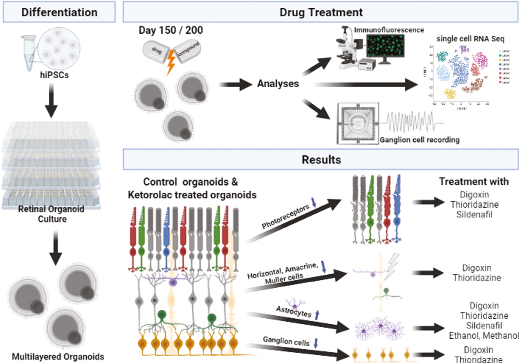
Retinal drug toxicity screening is essential for the development of safe treatment strategies for a large number of diseases. To this end, retinal organoids derived from human pluripotent stem cells (hPSCs) provide a suitable screening platform due to their similarity to the human retina and the ease of generation in large-scale formats. In this study, two hPSC cell lines were differentiated to retinal organoids, which comprised all key retinal cell types in multiple nuclear and synaptic layers. Single-cell RNA-Seq of retinal organoids indicated the maintenance of retinal ganglion cells and development of bipolar cells: both cell types segregated into several subtypes. Ketorolac, digoxin, thioridazine, sildenafil, ethanol, and methanol were selected as key compounds to screen on retinal organoids because of their well-known retinal toxicity profile described in the literature. Exposure of the hPSC-derived retinal organoids to digoxin, thioridazine, and sildenafil resulted in photoreceptor cell death, while digoxin and thioridazine additionally affected all other cell types, including Müller glia cells. All drug treatments caused activation of astrocytes, indicated by dendrites sprouting into neuroepithelium. The ability to respond to light was preserved in organoids although the number of responsive retinal ganglion cells decreased after drug exposure. These data indicate similar drug effects in organoids to those reported in in vivo models and/or in humans, thus providing the first robust experimental evidence of their suitability for toxicological studies.

Brain Slices
(paper)
Implementation of biohybrid olfactory bulb on a high-density CMOS-chip to reveal large-scale spatiotemporal circuit information
Biosens. Bioelectron. (2022). DOI: 10.1016/j.bios.2021.113834
2022
Keywords:
Large-scale multi-site biosensors are essential to probe the olfactory bulb (OB) circuitry for understanding the spatiotemporal dynamics of simultaneous discharge patterns. Current ex-vivo biosensing techniques are limited to recording a small set of neurons and cannot provide an adequate resolution, which hinders revealing the fast dynamic underlying the information coding mechanisms in the OB circuit. Here, we demonstrate a novel biohybrid OB-CMOS biosensing platform to decipher the cross-scale dynamics of the OB electrogenesis and quantify the distinct neuronal coding properties. The approach with 4096-microelectrodes offers a non-invasive, label-free, bioelectrical imaging to decode simultaneous firing patterns from thousands of connected neuronal ensembles in acute OB slices. The platform can measure spontaneous and drug-induced extracellular field potential activity with substantially improved spatiotemporal resolution over conventional OB-based biosensors. Also, we employ our OB-CMOS recordings to perform multidimensional analysis to instantiate specific neurophysiological metrics underlying the olfactory spatiotemporal coding that emerged from the OB interconnected layers. Our results delineate the computational implications of large-scale activity patterns in functional olfactory processing. The systematic interplay of the experimental CMOS-base platform architecture and the high-content characterization of the olfactory circuit with various computational analyses endow significant functional interrogations of the OB information processing, high-spatiotemporal connectivity mapping, and global circuit dynamics. Thus, our study can inspire the design of advanced biomimetic olfactory-based biosensors and neuromorphic approaches for diagnostic biomarkers and drug discovery applications.

Brain Slices
(paper)
Non-Linear Frequency Dependence of Neurovascular Coupling in the Cerebellar Cortex Implies Vasodilation–Vasoconstriction Competition
Cells (2022). DOI: 10.3390/cells11061047
2022
Keywords:

Neurovascular coupling (NVC) is the process associating local cerebral blood flow (CBF) to neuronal activity (NA). Although NVC provides the basis for the blood oxygen level dependent (BOLD) effect used in functional MRI (fMRI), the relationship between NVC and NA is still unclear. Since recent studies reported cerebellar non-linearities in BOLD signals during motor tasks execution, we investigated the NVC/NA relationship using a range of input frequencies in acute mouse cerebellar slices of vermis and hemisphere. The capillary diameter increased in response to mossy fiber activation in the 6–300 Hz range, with a marked inflection around 50 Hz (vermis) and 100 Hz (hemisphere). The corresponding NA was recorded using high-density multi-electrode arrays and correlated to capillary dynamics through a computational model dissecting the main components of granular layer activity. Here, NVC is known to involve a balance between the NMDAR-NO pathway driving vasodilation and the mGluRs-20HETE pathway driving vasoconstriction. Simulations showed that the NMDAR-mediated component of NA was sufficient to explain the time course of the capillary dilation but not its non-linear frequency dependence, suggesting that the mGluRs-20HETE pathway plays a role at intermediate frequencies. These parallel control pathways imply a vasodilation–vasoconstriction competition hypothesis that could adapt local hemodynamics at the microscale bearing implications for fMRI signals interpretation.

Brain Slices
(paper)
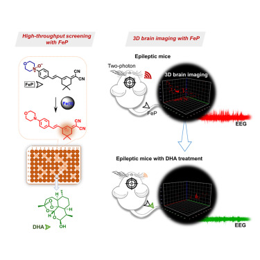
3D two-photon brain imaging reveals dihydroartemisinin exerts antiepileptic effects by modulating iron homeostasis
Cell Chemical Biology (2021). DOI: 10.1016/j.chembiol.2021.12.006
2021
Keywords:
Highlights
• FeP, a fluorescent probe, is suitable for brain imaging ferrous iron flux in vivo
• 3D two-photon imaging reveals elevation of ferrous iron in the epileptic mouse brain
• Dihydroartemisinin (DHA) can modulate iron homeostasis in the epileptic mouse
• DHA exhibits a potential antiepileptic effect in the epileptic mouse model
Summary
Imbalanced iron homeostasis plays a crucial role in neurological diseases, yet direct imaging evidence revealing the distribution of active ferrous iron (Fe2+) in the living brain remains scarce. Here, we present a near-infrared excited two-photon fluorescent probe (FeP) for imaging changes of Fe2+ flux in the living epileptic mouse brain. In vivo 3D two-photon brain imaging with FeP directly revealed abnormal elevation of Fe2+ in the epileptic mouse brain. Moreover, we found that dihydroartemisinin (DHA), a lead compound discovered through probe-based high-throughput screening, plays a critical role in modulating iron homeostasis. In addition, we revealed that DHA might exert its antiepileptic effects by modulating iron homeostasis in the brain and finally inhibiting ferroptosis. This work provides a reliable chemical tool for assessing the status of ferrous iron in the living epileptic mouse brain and may aid the rapid discovery of antiepileptic drug candidates.
Imbalanced iron homeostasis plays a crucial role in neurological diseases, yet direct imaging evidence revealing the distribution of active ferrous iron (Fe2+) in the living brain remains scarce. Here, we present a near-infrared excited two-photon fluorescent probe (FeP) for imaging changes of Fe2+ flux in the living epileptic mouse brain. In vivo 3D two-photon brain imaging with FeP directly revealed abnormal elevation of Fe2+ in the epileptic mouse brain. Moreover, we found that dihydroartemisinin (DHA), a lead compound discovered through probe-based high-throughput screening, plays a critical role in modulating iron homeostasis. In addition, we revealed that DHA might exert its antiepileptic effects by modulating iron homeostasis in the brain and finally inhibiting ferroptosis. This work provides a reliable chemical tool for assessing the status of ferrous iron in the living epileptic mouse brain and may aid the rapid discovery of antiepileptic drug candidates.

Cardiomyocyte
(paper)
All-Optical and Label-Free Stimulation of Action Potentials in Neurons and Cardiomyocytes by Plasmonic Porous Metamaterials
Adv. Sci. (2021). DOI: 10.1002/advs.202100627
2021
Keywords:
Optical stimulation technologies are gaining great consideration in cardiology, neuroscience studies, and drug discovery pathways by providing control over cell activity with high spatio-temporal resolution. However, this high precision requires manipulation of biological processes at genetic level concealing its development from broad scale application. Therefore, translating these technologies into tools for medical or pharmacological applications remains a challenge. Here, an all-optical nongenetic method for the modulation of electrogenic cells is introduced. It is demonstrated that plasmonic metamaterials can be used to elicit action potentials by converting near infrared laser pulses into stimulatory currents. The suggested approach allows for the stimulation of cardiomyocytes and neurons directly on commercial complementary metal-oxide semiconductor microelectrode arrays coupled with ultrafast pulsed laser, providing both stimulation and network-level recordings on the same device.

Brain Slices
(paper)
Arhgap22 Disruption Leads to RAC1 Hyperactivity Affecting Hippocampal Glutamatergic Synapses and Cognition in Mice
Mol. Neurobiol. (2021). DOI: 10.1007/s12035-021-02502-x
2021
Keywords:
Rho GTPases are a class of G-proteins involved in several aspects of cellular biology, including the regulation of actin cytoskeleton. The most studied members of this family are RHOA and RAC1 that act in concert to regulate actin dynamics. Recently, Rho GTPases gained much attention as synaptic regulators in the mammalian central nervous system (CNS). In this context, ARHGAP22 protein has been previously shown to specifically inhibit RAC1 activity thus standing as critical cytoskeleton regulator in cancer cell models; however, whether this function is maintained in neurons in the CNS is unknown. Here, we generated a knockout animal model for arhgap22 and provided evidence of its role in the hippocampus. Specifically, we found that ARHGAP22 absence leads to RAC1 hyperactivity and to an increase in dendritic spine density with defects in synaptic structure, molecular composition, and plasticity. Furthermore, arhgap22 silencing causes impairment in cognition and a reduction in anxiety-like behavior in mice. We also found that inhibiting RAC1 restored synaptic plasticity in ARHGAP22 KO mice. All together, these results shed light on the specific role of ARHGAP22 in hippocampal excitatory synapse formation and function as well as in learning and memory behaviors.
Organoids & Spheroids
(conf. proc.)
Human derived cortical excitatory neurospheroids showed spontaneous activity on micro electrodes array
NER 2021. DOI: 10.1109/NER49283.2021.9441261
2021
Keywords:
Human-induced pluripotent stem cells (hiPSCs) with their differentiation protocols, constitute a potential tool to investigate the various biological mechanisms of different human cells, such as those of the central nervous system. With the advent of such technique, we have increased the knowledge of biological mechanisms of neural diseases and, new therapies are now emerging. In particular, three-dimensional (3D) neural cell culture models including brain organoids and neurospheroids are increasingly used as in vitro platforms for studying human brain cell biology and drug screening, in genome engineering and transplantation as potential treatment for some neurodegenerative diseases. In this work, we exploited a particular differentiation protocol to generate engineered excitatory cortical neurospheroids of human origin. To assess functional network activity, we used standard Micro Electrodes Arrays (60 channels) and for the first time CMOS based devices (4096 channels). Sample cultures showed electrophysiological activity in 4 weeks and these first results suggest future possible applications for drug screening and transplantation.
.svg)


