


Neuronal Cultures
(paper)
A Monolayer System for the Efficient Generation of Motor Neuron Progenitors and Functional Motor Neurons from Human Pluripotent Stem Cells
Cells (2021). DOI: 10.3390/cells10051127

2021
Keywords:
Methods for the conversion of human induced pluripotent stem cells (hiPSCs) into motor neurons (MNs) have opened to the generation of patient-derived in vitro systems that can be exploited for MN disease modelling. However, the lack of simplified and consistent protocols and the fact that hiPSC-derived MNs are often functionally immature yet limit the opportunity to fully take advantage of this technology, especially in research aimed at revealing the disease phenotypes that are manifested in functionally mature cells. In this study, we present a robust, optimized monolayer procedure to rapidly convert hiPSCs into enriched populations of motor neuron progenitor cells (MNPCs) that can be further amplified to produce a large number of cells to cover many experimental needs. These MNPCs can be efficiently differentiated towards mature MNs exhibiting functional electrical and pharmacological neuronal properties. Finally, we report that MN cultures can be long-term maintained, thus offering the opportunity to study degenerative phenomena associated with pathologies involving MNs and their functional, networked activity. These results indicate that our optimized procedure enables the efficient and robust generation of large quantities of MNPCs and functional MNs, providing a valid tool for MNs disease modelling and for drug discovery applications.

Brain Slices
(paper)
Parp1 hyperactivity couples DNA breaks to aberrant neuronal calcium signalling and lethal seizures
EMBO Rep (2021). DOI: 10.15252/embr.202051851
2021
Keywords:
Defects in DNA single‐strand break repair (SSBR) are linked with neurological dysfunction but the underlying mechanisms remain poorly understood. Here, we show that hyperactivity of the DNA strand break sensor protein Parp1 in mice in which the central SSBR protein Xrcc1 is conditionally deleted (Xrcc1Nes‐Cre) results in lethal seizures and shortened lifespan. Using electrophysiological recording and synaptic imaging approaches, we demonstrate that aberrant Parp1 activation triggers seizure‐like activity in Xrcc1‐defective hippocampus ex vivo and deregulated presynaptic calcium signalling in isolated hippocampal neurons in vitro. Moreover, we show that these defects are prevented by Parp1 inhibition or deletion and, in the case of Parp1 deletion, that the lifespan of Xrcc1Nes‐Cre mice is greatly extended. This is the first demonstration that lethal seizures can be triggered by aberrant Parp1 activity at unrepaired SSBs, highlighting PARP inhibition as a possible therapeutic approach in hereditary neurological disease.

Cardiomyocyte
(paper)
Improving reliability and reducing costs of cardiotoxicity assessments using laser-induced cell poration on microelectrode arrays
Toxicol. Appl. Pharmacol. (2021). DOI: 10.1016/j.taap.2021.115480
2021
Keywords:

Drug-induced cardiotoxicity is a major barrier to drug development and a main cause of withdrawal of marketed drugs. Drugs can strongly alter the spontaneous functioning of the heart by interacting with the cardiac membrane ion channels. If these effects only surface during in vivo preclinical tests, clinical trials or worse after commercialization, the societal and economic burden will be significant and seriously hinder the efficient drug development process. Hence, cardiac safety pharmacology requires in vitro electrophysiological screening assays of all drug candidates to predict cardiotoxic effects before clinical trials. In the past 10 years, microelectrode array (MEA) technology began to be considered a valuable approach in pharmaceutical applications. However, an effective tool for high-throughput intracellular measurements, compatible with pharmaceutical standards, is not yet available. Here, we propose laser-induced optoacoustic poration combined with CMOS-MEA technology as a reliable and effective platform to detect cardiotoxicity. This approach enables the acquisition of high-quality action potential recordings from large numbers of cardiomyocytes within the same culture well, providing reliable data using single-well MEA devices and single cardiac syncytia per each drug. Thus, this technology could be applied in drug safety screening platforms reducing times and costs of cardiotoxicity assessments, while simultaneously improving the data reliability.

Brain Slices
(paper)
Sushi domain-containing protein 4 controls synaptic plasticity and motor learning
eLife (2021) DOI: 10.7554/eLife.65712
2021
Keywords:
Fine control of protein stoichiometry at synapses underlies brain function and plasticity. How proteostasis is controlled independently for each type of synaptic protein in a synapse-specific and activity-dependent manner remains unclear. Here, we show that Susd4, a gene coding for a complement-related transmembrane protein, is expressed by many neuronal populations starting at the time of synapse formation. Constitutive loss-of-function of Susd4 in the mouse impairs motor coordination adaptation and learning, prevents long-term depression at cerebellar synapses, and leads to misregulation of activity-dependent AMPA receptor subunit GluA2 degradation. We identified several proteins with known roles in the regulation of AMPA receptor turnover, in particular ubiquitin ligases of the NEDD4 subfamily, as SUSD4 binding partners. Our findings shed light on the potential role of SUSD4 mutations in neurodevelopmental diseases.

Acute Retina
(paper)
Transplanted pluripotent stem cell‐derived photoreceptor precursors elicit conventional and unusual light responses in mice with advanced retinal degeneration
Stem Cells Journal (2021). DOI: 10.1002/stem.3365
2021
Keywords:
Retinal dystrophies often lead to blindness. Developing therapeutic interventions to restore vision is therefore of paramount importance. Here we demonstrate the ability of pluripotent stem cell-derived cone precursors to engraft and restore light responses in the Pde6brd1 mouse, an end-stage photoreceptor degeneration model. Our data show that up to 1.5% of precursors integrate into the host retina, differentiate into cones, and engraft in close apposition to the host bipolar cells. Half of the transplanted mice exhibited visual behavior and of these 33% showed binocular light sensitivity. The majority of retinal ganglion cells exhibited contrast-sensitive ON, OFF or ON-OFF light responses and even motion sensitivity; however, quite a few exhibited unusual responses (eg, light-induced suppression), presumably reflecting remodeling of the neural retina. Our data indicate that despite relatively low engraftment yield, pluripotent stem cell-derived cone precursors can elicit light responsiveness even at advanced degeneration stages. Further work is needed to improve engraftment yield and counteract retinal remodeling to achieve useful clinical applications.

Acute Retina
(paper)
Modeling a population of retinal ganglion cells with restricted Boltzmann machines
Sci. Rep. (2020). DOI: 10.1038/s41598-020-73691-z
2020
Keywords:
The retina is a complex circuit of the central nervous system whose aim is to encode visual stimuli prior the higher order processing performed in the visual cortex. Due to the importance of its role, modeling the retina to advance in interpreting its spiking activity output is a well studied problem. In particular, it has been shown that latent variable models can be used to model the joint distribution of Retinal Ganglion Cells (RGCs). In this work, we validate the applicability of Restricted Boltzmann Machines to model the spiking activity responses of a large a population of RGCs recorded with high-resolution electrode arrays. In particular, we show that latent variables can encode modes in the RGC activity distribution that are closely related to the visual stimuli. In contrast to previous work, we further validate our findings by comparing results associated with recordings from retinas under normal and altered encoding conditions obtained by pharmacological manipulation. In these conditions, we observe that the model reflects well-known physiological behaviors of the retina. Finally, we show that we can also discover temporal patterns, associated with distinct dynamics of the stimuli.

Brain Slices
(paper)
Neuronal Glutamatergic Network Electrically Wired with Silent But Activatable Gap Junctions
The Journal of Neuroscience (2020). DOI: https://doi.org/10.1523/JNEUROSCI.2590-19.2020
2020
Keywords:
It is widely assumed that electrical synapses in the mammalian brain, especially between interneurons, underlie neuronal synchrony. In the hippocampus, principal cells also establish electrical synapses with each other and have also been implicated in network oscillations, whereby the origin of fast electrical activity has been attributed to ectopic spikelets and dendro-dendritic or axo-axonal gap junctions. However, if electrical synapses were in axo-dendritic connections, where chemical synapses occur, the synaptic events would be mixed, having an electrical component preceding the chemical one. This type of communication is less well studied, mainly because it is not easily detected. Moreover, a possible scenario could be that an electrical synapse coexisted with a chemical one, but in a nonconductive state; hence, it would be considered inexistent. Could chemical synapses have a quiescent electrical component? If so, can silent electrical synapses be activated to be detected? We addressed this possibility, and we here report that, indeed, the connexin-36-containing glutamatergic mossy fiber synapses of the rat hippocampus express previously unrecognized electrical synapses, which are normally silent. We reveal that these synapses are pH sensitive, actuate in vitro and in vivo, and that the electrical signaling is bidirectional. With the simultaneous recording of hundreds of cells, we could reveal the existence of an electrical circuit in the hippocampus of adult rats of either sex consisting of principal cells where the nodes are interregional glutamatergic synapses containing silent but ready-to-use gap junctions.

Organoids & Spheroids
(paper)
Room temperature shipment does not affect the biological activity of pluripotent stem cell derived retinal organoids
Plos One (2020). DOI: https://doi.org/10.1371/journal.pone.0233860
2020
Keywords:
The generation of laminated and light responsive retinal organoids from induced pluripotent stem cells (iPSCs) provides a powerful tool for the study of retinal diseases and drug discovery and a robust platform for cell-based therapies. The aim of this study is to investigate whether retinal organoids can retain their morphological and functional characteristics upon storage at room temperature (RT) conditions and shipment by air using a commercially available container that maintains the environment at ambient temperature. Morphological analysis and measurements of neuroepithelial thickness revealed no differences between control, RT incubated and shipped organoids. Similarly immunohistochemical analysis showed no differences in cell type composition and position within the laminated retinal structure. All groups showed a similar response to light, suggesting that the biological function of retinal organoids was not affected by RT storage or shipment. These findings provide an advance in transport of ready-made retinal organoids, increasing their availability to many research and pharma labs worldwide and facilitating cross-collaborative research.

Brain Slices
(paper)
A Claustrum in Reptiles and its role in Slow-Wave Sleep
Nature (2020). DOI: 10.1038/s41586-020-1993-6
2020
Keywords:
The mammalian claustrum, owing to its widespread connectivity with other forebrain structures, has been hypothesized to mediate functions that range from decision-making to consciousness1. Here we report that a homologue of the claustrum, identified by single-cell transcriptomics and viral tracing of connectivity, also exists in a reptile—the Australian bearded dragon Pogona vitticeps. In Pogona, the claustrum underlies the generation of sharp waves during slow-wave sleep. The sharp waves, together with superimposed high-frequency ripples2, propagate to the entire neighbouring pallial dorsal ventricular ridge (DVR). Unilateral or bilateral lesions of the claustrum suppress the production of sharp-wave ripples during slow-wave sleep in a unilateral or bilateral manner, respectively, but do not affect the regular and rapidly alternating sleep rhythm that is characteristic of sleep in this species3. The claustrum is thus not involved in the generation of the sleep rhythm itself. Tract tracing revealed that the reptilian claustrum projects widely to a variety of forebrain areas, including the cortex, and that it receives converging inputs from, among others, areas of the mid- and hindbrain that are known to be involved in wake–sleep control in mammals4,5,6. Periodically modulating the concentration of serotonin in the claustrum, for example, caused a matching modulation of sharp-wave production there and in the neighbouring DVR. Using transcriptomic approaches, we also identified a claustrum in the turtle Trachemys scripta, a distant reptilian relative of lizards. The claustrum is therefore an ancient structure that was probably already present in the brain of the common vertebrate ancestor of reptiles and mammals. It may have an important role in the control of brain states owing to the ascending input it receives from the mid- and hindbrain, its widespread projections to the forebrain and its role in sharp-wave generation during slow-wave sleep.
Acute Retina
(paper)
Electrical Coupling of Heterotypic Ganglion Cells in the Mammalian Retina
J. Neurosci. (2020). DOI: 10.1523/JNEUROSCI.1374-19.2019
2020
Keywords:
Electrical coupling has been reported to occur only between homotypic retinal ganglion cells, in line with the concept of parallel processing in the early visual system. Here, however, we show reciprocal correlated firing between heterotypic ganglion cells in multielectrode array recordings during light stimulation in retinas of adult guinea pigs of either sex. Heterotypic coupling was further confirmed via tracer spread after intracellular injections of single cells with neurobiotin. Both electrically coupled cell types were sustained ON center ganglion cells but showed distinct light response properties and receptive field sizes. We identified one of the involved cell types as sustained ON α-ganglion cells. The presence of electrical coupling between heterotypic ganglion cells introduces a network motif in which the signals of distinct ganglion cell types are partially mixed at the output stage of the retina.

Signal Processing
(paper)
Neural field models for latent state inference: Application to large-scale neuronal recordings
PLOS Computational Biology (2019). DOI: https://doi.org/10.1371/journal.pcbi.1007442
2019
Keywords:
Large-scale neural recording methods now allow us to observe large populations of identified single neurons simultaneously, opening a window into neural population dynamics in living organisms. However, distilling such large-scale recordings to build theories of emergent collective dynamics remains a fundamental statistical challenge. The neural field models of Wilson, Cowan, and colleagues remain the mainstay of mathematical population modeling owing to their interpretable, mechanistic parameters and amenability to mathematical analysis. Inspired by recent advances in biochemical modeling, we develop a method based on moment closure to interpret neural field models as latent state-space point-process models, making them amenable to statistical inference. With this approach we can infer the intrinsic states of neurons, such as active and refractory, solely from spiking activity in large populations. After validating this approach with synthetic data, we apply it to high-density recordings of spiking activity in the developing mouse retina. This confirms the essential role of a long lasting refractory state in shaping spatiotemporal properties of neonatal retinal waves. This conceptual and methodological advance opens up new theoretical connections between mathematical theory and point-process state-space models in neural data analysis.

Acute Retina
(paper)
Investigating the Effects of Mechanical Stimulation on Retinal Ganglion Cell Spontaneous Spiking Activity
Front. Cell. Neurosci. (2019). DOI: https://doi.org/10.3389/fnins.2019.01023
2019
Keywords:
Mechanical forces are increasingly recognized as major regulators of several physiological processes at both the molecular and cellular level; therefore, a deep understanding of the sensing of these forces and their conversion into electrical signals are essential for studying the mechanosensitive properties of soft biological tissues. To contribute to this field, we present a dual-purpose device able to mechanically stimulate retinal tissue and to record the spiking activity of retinal ganglion cells (RGCs). This new instrument relies on combining ferrule-top micro-indentation, which provides local measurements of viscoelasticity, with high-density multi-electrode array (HD-MEAs) to simultaneously record the spontaneous activity of the retina. In this paper, we introduce this instrument, describe its technical characteristics, and present a proof-of-concept experiment that shows how RGC spiking activity of explanted mice retinas respond to mechanical micro-stimulations of their photoreceptor layer. The data suggest that, under specific conditions of indentation, the retina perceive the mechanical stimulation as modulation of the visual input, besides the longer time-scale of activation, and the increase in spiking activity is not only localized under the indentation probe, but it propagates across the retinal tissue.

Neuronal Cultures
(conf. proc.)
Transfer Entropy Based Connectivity Estimation of Spontaneously Firing Hippocampal Cultures on Multi Electrode Arrays
International Joint Conference on Neural Networks (2019). DOI: 10.1109/IJCNN.2019.8851864
2019
Keywords:
Accurate estimation of interactions in neuronal circuits is critical in understanding neural information processing and the neuronal dynamics of emergent networks. Transfer entropy(TE) is a model-free information theoretic measure of flow of information between two random processes. TE has recently gained much popularity due to its effectiveness in estimating effective connectivity among neurons from simulated networks. However, experimental recordings inherently lack the ground truth information of neural connectivity - making it difficult to identify true connections from spurious connections. To tackle this problem, we present a superimposition method where the estimated connectivity is superimposed onto the spatial firing density plot. The firing density plot, on itself, does not provide connectivity information but we assume that frequently firing channels are more likely to have more active interactions. A neuron's firing depends on the presynaptic inputs, and highly interconnected neurons tend to have more presynaptic inputs resulting in more frequent firing. The firing density plot is organised in a spatial layout, which corresponds substantially to the structural layout. We demonstrated that the connectivity estimated closely corresponds to the firing density plot when superimposed onto the firing density plot. To strengthen the arguement, we analyse the firing count and the connectivity inferred for each randomly sampled channel and found a positively correlated relationship.
Organoids & Spheroids
(paper)
Systematic Comparison of Retinal Organoid Differentiation from Human Pluripotent Stem Cells Reveals Stage Specific, Cell Line, and Methodological Differences
Stem Cells Transl. Med. (2019), DOI: 10.1002/sctm.18-0267
2019
Keywords:
A major goal in the stem cell field is to generate tissues that can be utilized as a universal tool for in vitro models of development and disease, drug development, or as a resource for patients suffering from disease or injury. Great efforts are being made to differentiate human pluripotent stem cells in vitro toward retinal tissue, which is akin to native human retina in its cytoarchitecture and function, yet the numerous existing retinal induction protocols remain variable in their efficiency and do not routinely produce morphologically or functionally mature photoreceptors. Herein, we determine the impact that the method of embryoid body (EB) formation and maintenance as well as cell line background has on retinal organoid differentiation from human embryonic stem cells and human induced pluripotent stem cells. Our data indicate that cell line-specific differences dominate the variables that underline the differentiation efficiency in the early stages of differentiation. In contrast, the EB generation method and maintenance conditions determine the later differentiation and maturation of retinal organoids. Of the latter, the mechanical method of EB generation under static conditions, accompanied by media supplementation with Y27632 for the first 48 hours of differentiation, results in the most consistent formation of laminated retinal neuroepithelium containing mature and electrophysiologically responsive photoreceptors. Collectively, our data provide substantive evidence for stage-specific differences in the ability to give rise to laminated retinae, which is determined by cell line-specific differences in the early stages of differentiation and EB generation/organoid maintenance methods at later stages.

Organoids & Spheroids
(paper)
Decellularised extracellular matrix-derived peptides from neural retina and retinal pigment epithelium enhance the expression of synaptic markers and light responsiveness of human pluripotent stem cell derived retinal organoids
Biomater. (2019) DOI: 10.1016/j.biomaterials.2019.01.028
2019
Keywords:

Tissue specific extracellular matrices (ECM) provide structural support and enable access to molecular signals and metabolites, which are essential for directing stem cell renewal and differentiation. To mimic this phenomenon in vitro, tissue decellularisation approaches have been developed, resulting in the generation of natural ECM scaffolds that have comparable physical and biochemical properties of the natural tissues and are currently gaining traction in tissue engineering and regenerative therapies due to the ease of standardised production, and constant availability. In this manuscript we report the successful generation of decellularised ECM-derived peptides from neural retina (decel NR) and retinal pigment epithelium (decel RPE), and their impact on differentiation of human pluripotent stem cells (hPSCs) to retinal organoids. We show that culture media supplementation with decel RPE and RPE-conditioned media (CM RPE) significantly increases the generation of rod photoreceptors, whilst addition of decel NR and decel RPE significantly enhances ribbon synapse marker expression and the light responsiveness of retinal organoids. Photoreceptor maturation, formation of correct synapses between retinal cells and recording of robust light responses from hPSC-derived retinal organoids remain unresolved challenges for the field of regenerative medicine. Enhanced rod photoreceptor differentiation, synaptogenesis and light response in response to addition of decellularised matrices from RPE and neural retina as shown herein provide a novel and substantial advance in generation of retinal organoids for drug screening, tissue engineering and regenerative medicine.
Acute Retina
(paper)
Non-parametric Physiological Classification of Retinal Ganglion Cells in the Mouse Retina
Front. Cell. Neurosci. (2018). DOI: https://doi.org/10.3389/fncel.2018.00481
2018
Keywords:
Retinal ganglion cells, the sole output neurons of the retina, exhibit surprising diversity. A recent study reported over 30 distinct types in the mouse retina, indicating that the processing of visual information is highly parallelised in the brain. The advent of high density multi-electrode arrays now enables recording from many hundreds to thousands of neurons from a single retina. Here we describe a method for the automatic classification of large-scale retinal recordings using a simple stimulus paradigm and a spike train distance measure as a clustering metric. We evaluate our approach using synthetic spike trains, and demonstrate that major known cell types are identified in high-density recording sessions from the mouse retina with around 1,000 retinal ganglion cells. A comparison across different retinas reveals substantial variability between preparations, suggesting pooling data across retinas should be approached with caution. As a parameter-free method, our approach is broadly applicable for cellular physiological classification in all sensory modalities.

Brain Slices
(conf. proc.)
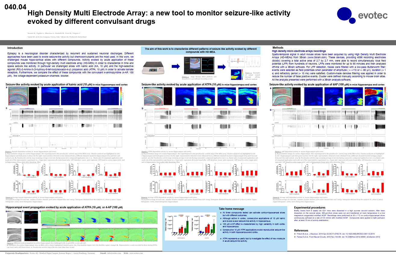
High Density Multi Electrode Array: a new tool to monitor seizure-like activity evoked by different convulsant drugs
SFN (2018). San Diego, CA, USA.
2018
Keywords:
Brain Slices
(paper)
Early Appearance and Spread of Fast Ripples in the Hippocampus in a Model of Cortical Traumatic Brain Injury
J. Neurosci. (2018). DOI: 10.1523/JNEUROSCI.3507-17.2018
2018
Keywords:
Fast ripples (FRs; activity of >250 Hz) have been considered as a biomarker of epileptic activity in the hippocampus and entorhinal cortex; it is thought that they signal the focus of seizure generation. Similar high-frequency network activity has been produced in vitro by changing extracellular medium composition, by using pro-epileptic substances, or by electrical stimulation. Here we study the propagation of these events between different subregions of the male rat hippocampus in a recently introduced experimental model of FRs in entorhinal cortex–hippocampal slices in vitro. By using a matrix of 4096 microelectrodes, the sites of initiation, propagation pathways, and spatiotemporal characteristics of activity patterns could be studied with unprecedented high resolution. To this end, we developed an analytic tool based on bidimensional current source density estimation, which delimits sinks and sources with a high precision and evaluates their trajectories using the concept of center of mass. With this methodology, we found that FRs can arise almost simultaneously at noncontiguous sites in the CA3-to-CA1 direction, underlying the spatial heterogeneity of epileptogenic foci, while continuous somatodendritic waves of activity develop. An unexpected, yet important propagation route is the propagation of activity from CA3 into the hilus and dentate gyrus. This pathway may cause reverberating activation of both regions, supporting sustained pathological network events and altered information processing in hippocampal networks.

Organoids & Spheroids
(paper)
Disrupted alternative splicing for genes implicated in splicing and ciliogenesis causes PRPF31 retinitis pigmentosa
Nat. Commun. (2018). DOI: 10.1038/s41467-018-06448-y
2018
Keywords:
Mutations in pre-mRNA processing factors (PRPFs) cause autosomal-dominant retinitis pigmentosa (RP), but it is unclear why mutations in ubiquitously expressed genes cause non-syndromic retinal disease. Here, we generate transcriptome profiles from RP11 (PRPF31-mutated) patient-derived retinal organoids and retinal pigment epithelium (RPE), as well as Prpf31+/− mouse tissues, which revealed that disrupted alternative splicing occurred for specific splicing programmes. Mis-splicing of genes encoding pre-mRNA splicing proteins was limited to patient-specific retinal cells and Prpf31+/− mouse retinae and RPE. Mis-splicing of genes implicated in ciliogenesis and cellular adhesion was associated with severe RPE defects that include disrupted apical – basal polarity, reduced trans-epithelial resistance and phagocytic capacity, and decreased cilia length and incidence. Disrupted cilia morphology also occurred in patient-derived photoreceptors, associated with progressive degeneration and cellular stress. In situ gene editing of a pathogenic mutation rescued protein expression and key cellular phenotypes in RPE and photoreceptors, providing proof of concept for future therapeutic strategies.
Signal Processing
(paper)
Identification of excitatory-inhibitory links and network topology in large-scale neuronal assemblies from multi-electrode recordings
PLOS Computational Biology (2018). DOI: https://doi.org/10.1371/journal.pcbi.1006381
2018
Keywords:
Functional-effective connectivity and network topology are nowadays key issues for studying brain physiological functions and pathologies. Inferring neuronal connectivity from electrophysiological recordings presents open challenges and unsolved problems. In this work, we present a cross-correlation based method for reliably estimating not only excitatory but also inhibitory links, by analyzing multi-unit spike activity from large-scale neuronal networks. The method is validated by means of realistic simulations of large-scale neuronal populations. New results related to functional connectivity estimation and network topology identification obtained by experimental electrophysiological recordings from high-density and large-scale (i.e., 4096 electrodes) microtransducer arrays coupled to in vitro neural populations are presented. Specifically, we show that: (i) functional inhibitory connections are accurately identified in in vitro cortical networks, providing that a reasonable firing rate and recording length are achieved; (ii) small-world topology, with scale-free and rich-club features are reliably obtained, on condition that a minimum number of active recording sites are available. The method and procedure can be directly extended and applied to in vivo multi-units brain activity recordings.

Cardiomyocyte
(paper)
Plasmonic meta-electrodes allow intracellular recordings at network level on high-density CMOS-multi-electrode arrays
Nat. Nanotechnol. (2018). DOI: https://doi.org/10.1038/s41565-018-0222-z
2018
Keywords:
The ability to monitor electrogenic cells accurately plays a pivotal role in neuroscience, cardiology and cell biology. Despite pioneering research and long-lasting efforts, the existing methods for intracellular recording of action potentials on the large network scale suffer limitations that prevent their widespread use. Here, we introduce the concept of a meta-electrode, a planar porous electrode that mimics the optical and biological behaviour of three-dimensional plasmonic antennas but also preserves the ability to work as an electrode. Its synergistic combination with plasmonic optoacoustic poration allows commercial complementary metal–oxide semiconductor multi-electrode arrays to record intracellular action potentials in large cellular networks. We apply this approach to measure signals from human-induced pluripotent stem cell-derived cardiac cells, rodent primary cardiomyocytes and immortalized cell types and demonstrate the possibility of non-invasively testing a variety of relevant drugs. Due to its robustness and easiness of use, we expect the method will be rapidly adopted by the scientific community and by pharmaceutical companies.
Technology
(paper)
Exploiting All Programmable SoCs in Neural Signal Analysis: A Closed-Loop Control for Large-Scale CMOS Multielectrode Arrays
IEEE Trans. Biomed. Circuits Syst. (2018). DOI: 10.1109/TBCAS.2018.2830659
2018
Keywords:
Microelectrode array (MEA) systems with up to several thousands of recording electrodes and electrical or optical stimulation capabilities are commercially available or described in the literature. By exploiting their submillisecond and micrometric temporal and spatial resolutions to record bioelectrical signals, such emerging MEA systems are increasingly used in neuroscience to study the complex dynamics of neuronal networks and brain circuits. However, they typically lack the capability of implementing real-time feedback between the detection of neuronal spiking events and stimulation, thus restricting large-scale neural interfacing to open-loop conditions. In order to exploit the potential of such large-scale recording systems and stimulation, we designed and validated a fully reconfigurable FPGA-based processing system for closed-loop multichannel control. By adopting a Xilinx Zynq-all-programmable system on chip that integrates reconfigurable logic and a dual-core ARM-based processor on the same device, the proposed platform permits low-latency preprocessing (filtering and detection) of spikes acquired simultaneously from several thousands of electrode sites. To demonstrate the proposed platform, we tested its performances through ex vivo experiments on the mice retina using a state-of-the-art planar high-density MEA that samples 4096 electrodes at 18 kHz and record light-evoked spikes from several thousands of retinal ganglion cells simultaneously. Results demonstrate that the platform is able to provide a total latency from whole-array data acquisition to stimulus generation below 2 ms. This opens the opportunity to design closed-loop experiments on neural systems and biomedical applications using emerging generations of planar or implantable large-scale MEA systems.
Organoids & Spheroids
(paper)
Human‐Induced Pluripotent Stem Cells Generate Light Responsive Retinal Organoids with Variable and Nutrient‐Dependent Efficiency
Stem Cells (2018). DOI: 10.1002/stem.2883
2018
Keywords:
The availability of in vitro models of the human retina in which to perform pharmacological and toxicological studies is an urgent and unmet need. An essential step for developing in vitro models of human retina is the ability to generate laminated, physiologically functional, and light-responsive retinal organoids from renewable and patient specific sources. We investigated five different human-induced pluripotent stem cell (iPSC) lines and showed a significant variability in their efficiency to generate retinal organoids. Despite this variability, by month 5 of differentiation, all iPSC-derived retinal organoids were able to generate light responses, albeit immature, comparable to the earliest light responses recorded from the neonatal mouse retina, close to the period of eye opening. All iPSC-derived retinal organoids exhibited at this time a well-formed outer nuclear like layer containing photoreceptors with inner segments, connecting cilium, and outer like segments. The differentiation process was highly dependent on seeding cell density and nutrient availability determined by factorial experimental design. We adopted the differentiation protocol to a multiwell plate format, which enhanced generation of retinal organoids with retinal-pigmented epithelium (RPE) and improved ganglion cell development and the response to physiological stimuli. We tested the response of iPSC-derived retinal organoids to Moxifloxacin and showed that similarly to in vivo adult mouse retina, the primary affected cell types were photoreceptors. Together our data indicate that light responsive retinal organoids derived from carefully selected and differentiation efficient iPSC lines can be generated at the scale needed for pharmacology and drug screening purposes.
Signal Processing
(conf. proc.)
Spike Train Synchrony Analysis of Neuronal Cultures
International Joint Conference on Neural Networks (IJCNN) (2018). DOI: 10.1109/IJCNN.2018.8489728
2018
Keywords:
Spike train synchrony estimation of neuronal cultures provides valuable insights into firing patterns of neurons in terms of degree of similarity or dissimilarity. These estimations have proven to be a useful tool in neuroscience since synchrony in neuronal networks is thought to be related to cognitive processes, sensory awareness, learning and neurological disorders. Many mathematical measures have been developed to quantify the degree of synchrony. These synchrony metrics are generally used for smaller sets of spike trains and not been explored for larger High Density Multi Electrode Arrays (HD-MEA) datasets with thousands of channels. Here, bivariate and multivariate ISI-distance and SPIKE-distance metrics are utilized on both synthetic and experimental HD-MEA datasets to quantify spike train synchrony. It is demonstrated that, despite the significant size of the datasets, the approaches are effective in identifying and quantifying interesting bursting or change in spike train behaviours which are not always obvious from the raster plot.

Brain Slices
(conf. proc.)
Purkinje cells firing recorded by a high density multi-electrode array: a new tool for compounds validation
FENS conference (2018). Berlin, Germany.
2018
Keywords:

.svg)


