


Neuronal Cultures
(paper)
Sloppiness in Spontaneously Active Neuronal Networks
J. Neurosci. (2015). DOI: 10.1523/JNEUROSCI.4421-14.2015.

2015
Keywords:
Various plasticity mechanisms, including experience-dependent, spontaneous, as well as homeostatic ones, continuously remodel neural circuits. Yet, despite fluctuations in the properties of single neurons and synapses, the behavior and function of neuronal assemblies are generally found to be very stable over time. This raises the important question of how plasticity is coordinated across the network. To address this, we investigated the stability of network activity in cultured rat hippocampal neurons recorded with high-density multielectrode arrays over several days. We used parametric models to characterize multineuron activity patterns and analyzed their sensitivity to changes. We found that the models exhibited sloppiness, a property where the model behavior is insensitive to changes in many parameter combinations, but very sensitive to a few. The activity of neurons with sloppy parameters showed faster and larger fluctuations than the activity of a small subset of neurons associated with sensitive parameters. Furthermore, parameter sensitivity was highly correlated with firing rates. Finally, we tested our observations from cell cultures on an in vivo recording from monkey visual cortex and we confirm that spontaneous cortical activity also shows hallmarks of sloppy behavior and firing rate dependence. Our findings suggest that a small subnetwork of highly active and stable neurons supports group stability, and that this endows neuronal networks with the flexibility to continuously remodel without compromising stability and function.

Neuronal Cultures
(paper)
Functional Connectivity Estimation over Large Networks at Cellular Resolution based on Electrophysiological Recordings and Structural Prior
Front. Neuroanat. (2014), DOI: 10.3389/fnana.2014.00137.
2014
Keywords:
Despite many structural and functional aspects of the brain organization have been extensively studied in neuroscience, we are still far from a clear understanding of the intricate structure-function interactions occurring in the multi-layered brain architecture, where billions of different neurons are involved. Although structure and function can individually convey a large amount of information, only a combined study of these two aspects can probably shade light on how brain circuits develop and operate at the cellular scale. Here, we propose a novel approach for refining functional connectivity estimates within neuronal networks using the structural connectivity as prior. This is done at the mesoscale, dealing with thousands of neurons while reaching, at the microscale, an unprecedented cellular resolution. The High-Density Micro Electrode Array (HD-MEA) technology, combined with fluorescence microscopy, offers the unique opportunity to acquire structural and functional data from large neuronal cultures approaching the granularity of the single cell. In this work, an advanced method based on probabilistic directional features and heat propagation is introduced to estimate the structural connectivity from the fluorescence image while functional connectivity graphs are obtained from the cross-correlation analysis of the spiking activity. Structural and functional information are then integrated by reweighting the functional connectivity graph based on the structural prior. Results show that the resulting functional connectivity estimates are more coherent with the network topology, as compared to standard measures purely based on cross-correlations and spatio-temporal filters. We finally use the obtained results to gain some insights on which features of the functional activity are more relevant to characterize actual neuronal interactions.

Technology
(conf. proc.)
Large-Scale recording of light-evoked responses in the retinal ganglion cell layer of the explanted retina: A new HD experimental platform
SFN November (2014). Washington, DC, USA.
2014
Keywords:
Aims: We introduce an innovative setup to simultaneously evoke and record in-vitro extracellular visual responses from Retinal Ganglion Cells (RGCs) with micrometer/cellular resolution from a complete whole-mount mouse retina. We show the capabilities of this setup by investigating the effects of the extra-classical receptive field of RGCs. The applicability of this setup ranges from the study of a complete biological system, to optogenetic experiments that may benefit from the precise control of a light stimulus (structured or not) and from the ability to simultaneously record spikes from thousands of single cells. Methods: Two main parts constitute the setup: the optical stimulator and the recording system. The recording system is a customized version of the commercially available Biocam adapted to fit the optical stimulator geometrical constraints. Briefly, the system is composed by a CMOS 4096-electrode array chip (electrode size 21 µm x 21 µm, inter-electrode separation 21 µm), mounted on hardware for real-time filtering and simultaneous recording of all the electrodes at a sampling rate of about 7KHz/channel. With an active area of 7.12 mm2 the system allows pan retinal recording from mouse explanted retina. Since the chip is not transparent, visual stimuli are delivered from the top of the chip by means of a DLP-projector, with 3-axis of free-movement, aligned with a single-convex lens that focuses the image on the biological sample. A beam-splitter, positioned in the optic pathway, direct the reflected light to a camera allowing to precisely align the stimulus with the recording area. To precisely control and synchronize the timing of the photo-stimulation with the data acquisition, custom hardware and software tools were developed. These also allow generation of complex photo-stimulation protocols, and to record the timestamp of the photo-stimulations with millisecond precision. Results: Here we show that a visual stimulus confined to a small region of the retina does affect the spontaneous activity in parts of the retina not exposed to the stimulus. In particular, spontaneous activity is reduced in the area not exposed to the stimulus for during stimulus presentation, and increases sharply and transiently at stimulus offset. Conclusions: Our integrated platform enables successful recording of a large number of RGCs in the mouse retina during photo stimulation. Our results show that such dense and large-scale recordings can reveal spatial processing mechanisms in the retina that are difficult to characterize using conventional technology.

Technology
(book chapter)
Integration of microstructured scaffolds, neurons, and multielectrode arrays
Progress in Brain Research (Vol. 214, pp. 415-442). Elsevier (2014). DOI: 10.1016/B978-0-444-63486-3.00017-7
2014
Keywords:
Recent progresses in neuroelectronics and lab-on-a-chip technologies are providing novel opportunities for neuroscience research and applications. However, the experimental performances of these novel devices are not only the result of the artificially implemented features, such as those resulting from advanced electrode materials, from electrode morphologies, or from the low noise levels of the front-end electronic circuits. Rather, these performances also strictly relay on the bioartificial interface established by neurons on these devices. Here, we focus on cell culture systems adapted to neuroelectronic devices that were developed for organizing and growing neural networks in two or three dimensions. These developments span the fields of biosensors, engineering, neuroscience, and novel nanostructures and materials. Additionally, they are at the origin of novel neuroartificial hybrid technologies that can be applied for the study of neuronal networks at unprecedented scales and for applications in neuroscience that use scaffolding micro-/nanostructures, neurons, and biomolecules for advanced neuroelectronic interfaces and novel cell culture systems.
Signal Processing
(conf. proc.)
Characterizing neural population response with high-density multielectrode arrays
SFN conference (2014). Washington, DC, USA.
2014
Keywords:


Technology
(conf. proc.)
Novel 3D plasmonic nano-electrodes for cellular investigations and neural interfaces
SPIE 9166, Biosensing and Nanomedicine VII (2014). DOI: 10.1117/12.2066274
2014
Keywords:
We propose the development of an innovative plasmonic-electronic multifunctional platform, capable at the same time of performing chemical analysis and electronic recordings from a cellular interface. The system, based on 3D hollow metallic nanotubes, integrated on customized multi-electrode-arrays, allows the study of neuronal signaling over different lengths, spanning from the molecular, to the cellular, to the network scale. Here we show that the same structures are efficient electric field enhancers, despite the continuous metal layer at the base, which connects them to the electric components of the integrated circuits. The methodology we propose, due to its simplicity and high throughput, has the potential for further improvements both in the field of plasmonics, and in the integration on large areas of commercial active electronic devices.

Neuronal Cultures
(conf. proc.)
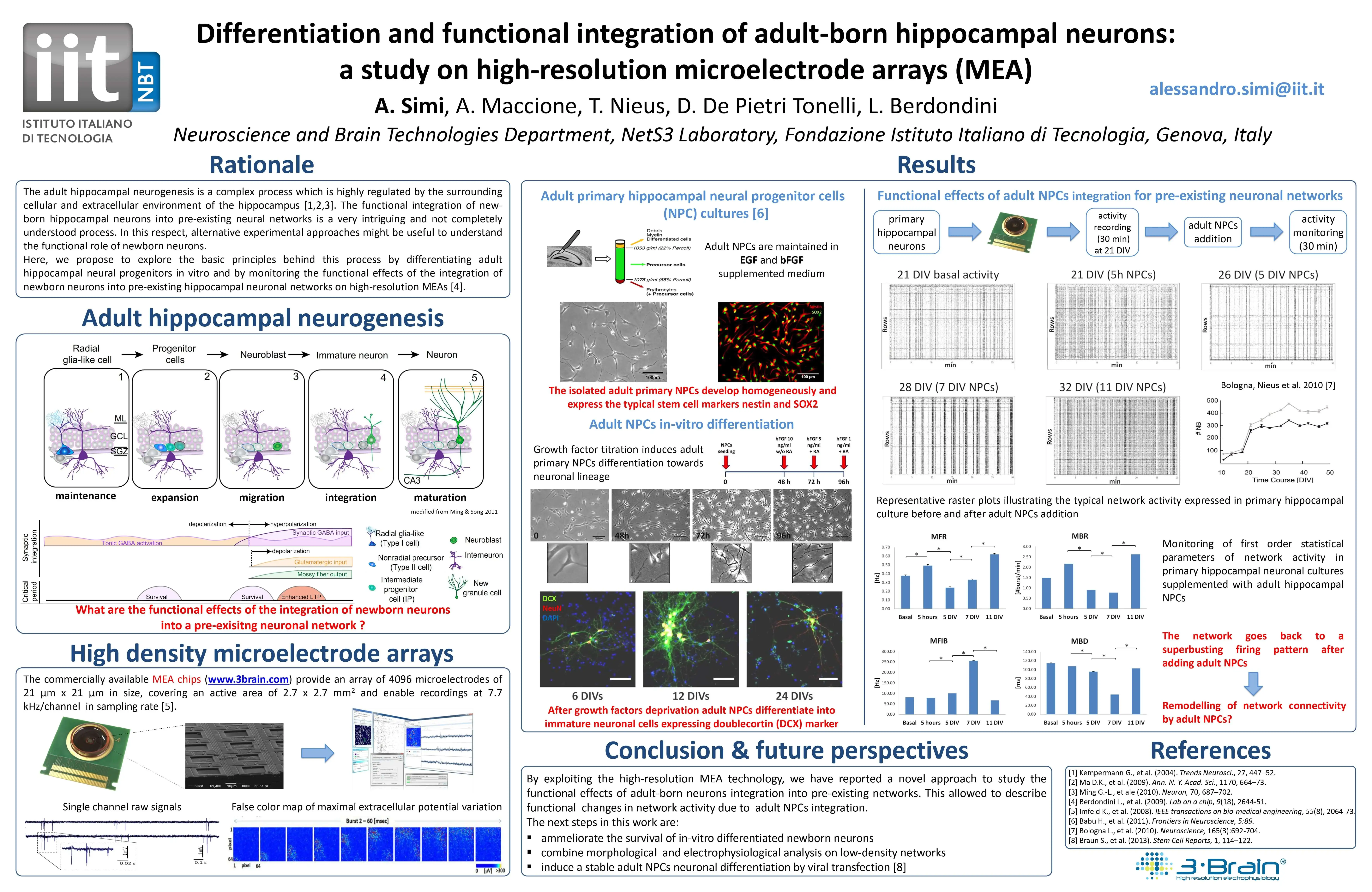
Differentiation and functional integration of adult-born hippocampal neurons: a study on high-resolution microelectrode arrays (MEA)
9th Federation of European Neuroscience Societies Meeting (FENS2014). Milan, Italy.
2014
Keywords:
Neuronal Cultures
(conf. proc.)
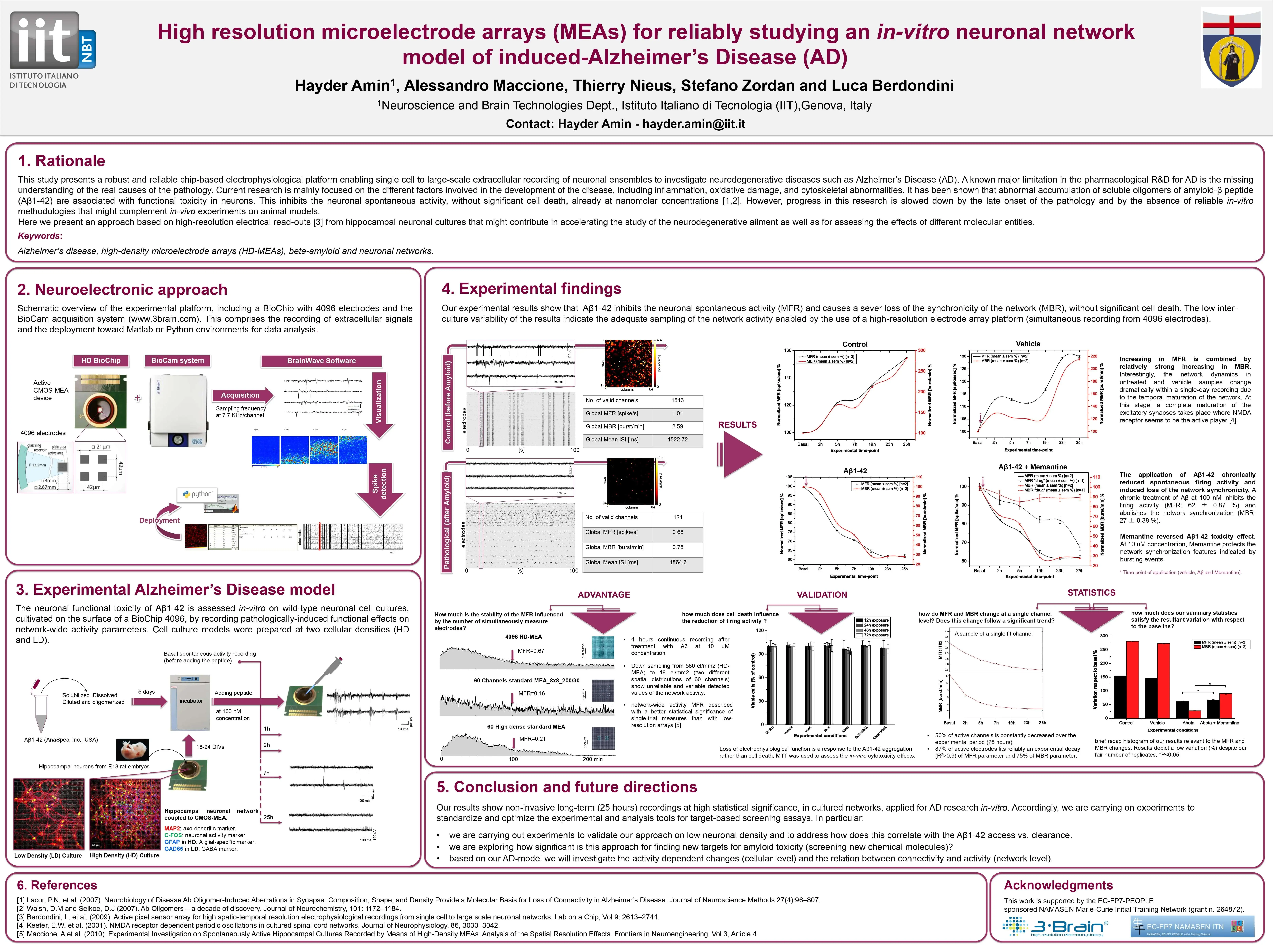
Alzheimer's disease (AD) in-vitro model: a novel drug screening approach on 4096-microelectrode recording arrays
9th Federation of European Neuroscience Societies Meeting (FENS2014). Milan, Italy.
2014
Keywords:

Acute Retina
(conf. proc.)
High Resolution Large-Scale Recordings of Light Responses from Mouse Retinal Ganglion Cells
MEA meeting (2014). Reutlingen, Germany.
2014
Keywords:
.webp)

Brain Slices
(paper)
Asynchronous GABA Release Is a Key Determinant of Tonic Inhibition and Controls Neuronal Excitability: A Study in the Synapsin II-/- Mouse
Cereb. Cortex (2014). DOI: 10.1093/cercor/bhu141
2014
Keywords:
Idiopathic epilepsies have frequently been linked to mutations in voltage-gated channels (channelopathies); recently, mutations in several genes encoding presynaptic proteins have been shown to cause epilepsy in humans and mice, indicating that epilepsy can also be considered a synaptopathy. However, the functional mechanisms by which presynaptic dysfunctions lead to hyperexcitability and seizures are not well understood. We show that deletion of synapsin II (Syn II), a presynaptic protein contributing to epilepsy predisposition in humans, leads to a loss of tonic inhibition in mouse hippocampal slices due to a dramatic decrease in presynaptic asynchronous GABA release. We also show that the asynchronous GABA release reduces postsynaptic cell firing, and the parallel impairment of asynchronous GABA release and tonic inhibition results in an increased excitability at both single-neuron and network levels. Restoring tonic inhibition with THIP (4,5,6,7-tetrahydroisoxazolo[5,4-c]pyridin-3-ol; gaboxadol), a selective agonist of δ subunit-containing GABAA receptors, fully rescues the SynII−/− epileptic phenotype both ex vivo and in vivo. The results demonstrate a causal relationship between the dynamics of GABA release and the generation of tonic inhibition, and identify a novel mechanism of epileptogenesis generated by dysfunctions in the dynamics of release that can be effectively targeted by novel antiepileptic strategies.

Technology
(conf. proc.)
3D plasmonic hollow nanoantennas as tools for neuroscience applications
CLEO: Science and Innovations (2014). DOI: 10.1364/CLEO_SI.2014.STh4H.3
2014
Keywords:
3D plasmonic nanoantennas were fabricated on active biodevices for in-vitro neuroscience experiments. The technique consents to realize nanoantennas patterns suitable for neurons culture and that can be used concurrently as intracellular nanoelectrodes and spectroscopic probes.

Neuronal Cultures
(conf. proc.)
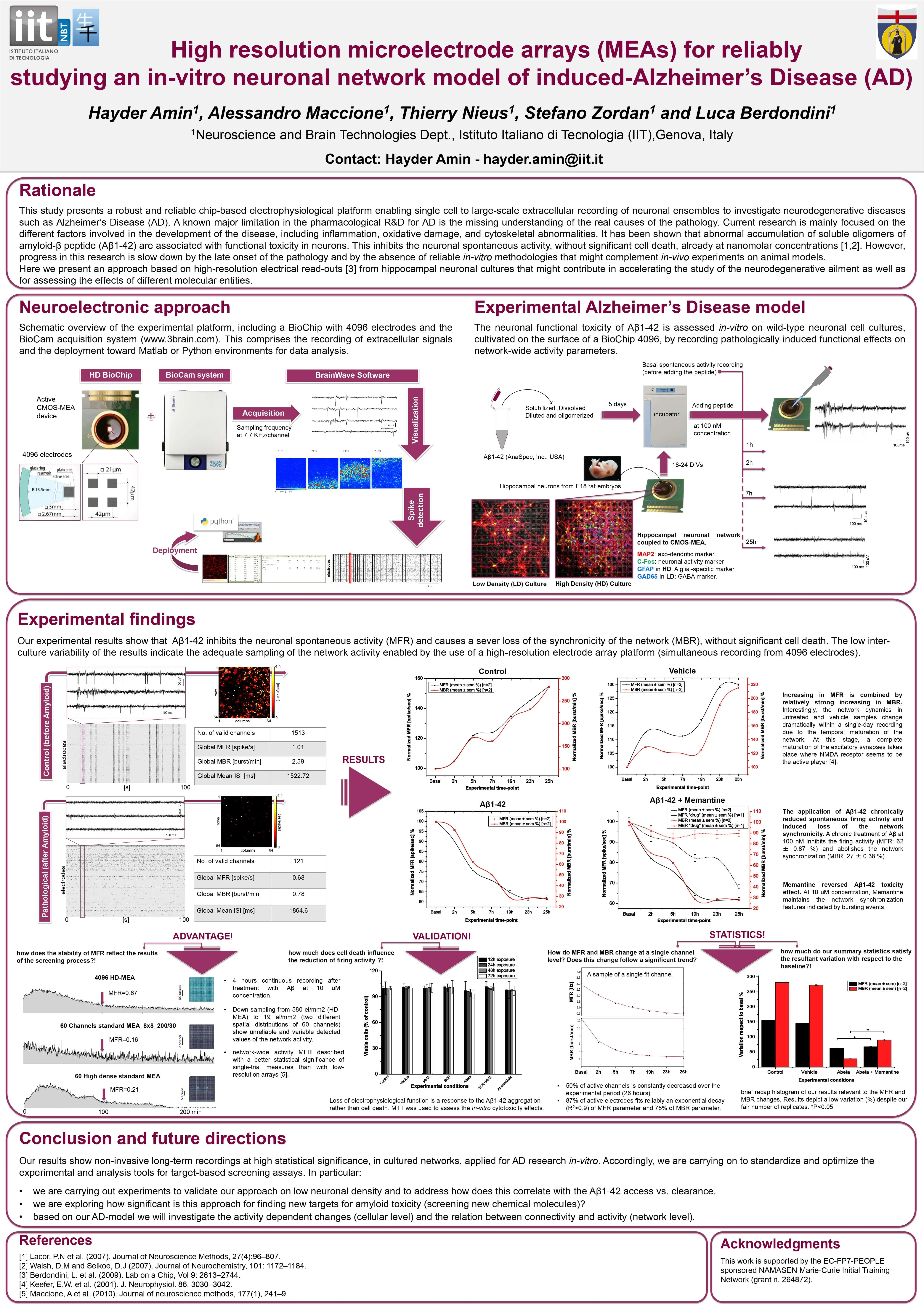
High resolution microelectrode arrays (MEAs) for reliably studying an in-vitro neuronal network model of induced-Alzheimer's disease
24th Anniversary World Congress on Biosensors (2014). Melbourne, Australia.
2014
Keywords:

Neuronal Cultures
(paper)
Dominant β-catenin mutations cause intellectual disability with recognizable syndromic features
The Journal of Clinical Investigation DOI: 10.1172/JCI70372
2024
Keywords:
The recent identification of multiple dominant mutations in the gene encoding β-catenin in both humans and mice has enabled exploration of the molecular and cellular basis of β-catenin function in cognitive impairment. In humans, β-catenin mutations that cause a spectrum of neurodevelopmental disorders have been identified. We identified de novo β-catenin mutations in patients with intellectual disability, carefully characterized their phenotypes, and were able to define a recognizable intellectual disability syndrome. In parallel, characterization of a chemically mutagenized mouse line that displays features similar to those of human patients with β-catenin mutations enabled us to investigate the consequences of β-catenin dysfunction through development and into adulthood. The mouse mutant, designated batface (Bfc), carries a Thr653Lys substitution in the C-terminal armadillo repeat of β-catenin and displayed a reduced affinity for membrane-associated cadherins. In association with this decreased cadherin interaction, we found that the mutation results in decreased intrahemispheric connections, with deficits in dendritic branching, long-term potentiation, and cognitive function. Our study provides in vivo evidence that dominant mutations in β-catenin underlie losses in its adhesion-related functions, which leads to severe consequences, including intellectual disability, childhood hypotonia, progressive spasticity of lower limbs, and abnormal craniofacial features in adults.

Acute Retina
(paper)
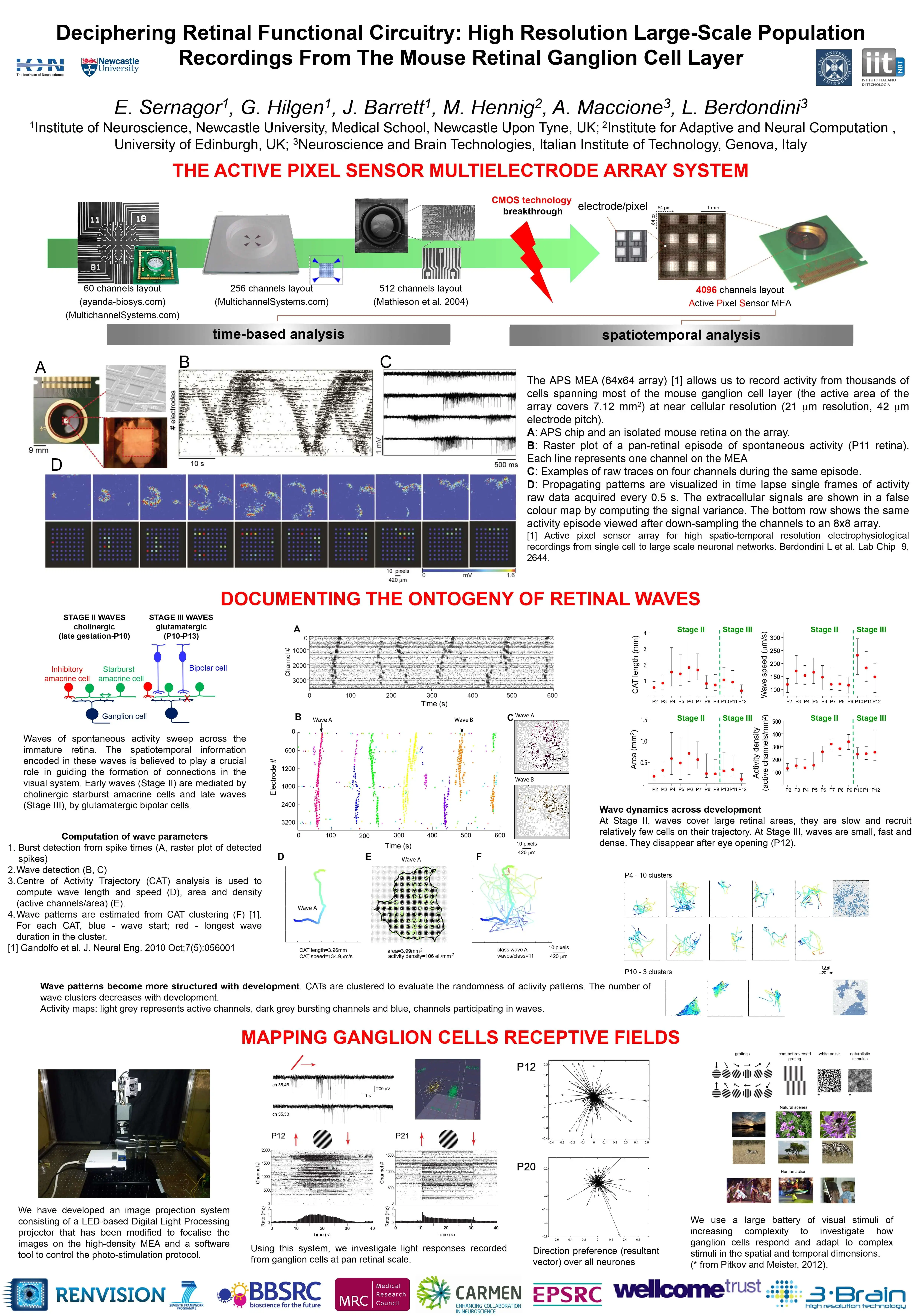
Following the Ontogeny of Retinal Waves: Pan-Retinal Recordings of Population Dynamics in the Neonatal Mouse
J. Physiol. (2014). DOI: 10.1113/jphysiol.2013.262840.
2014
Keywords:
Key points
- Novel pan-retinal recordings of mouse retinal waves were obtained at near cellular resolution using a large-scale, high-density array of 4096 electrodes to investigate changes in wave spatiotemporal properties from postnatal day 2 to eye opening.
- Early cholinergic waves are large, slow and random, with low cellular recruitment.
- A developmental shift in GABAA signalling from depolarizing to hyperpolarizing influences the dynamics of cholinergic waves.
- Glutamatergic waves that occur just before eye opening are focused, faster, denser, non-random and repetitive.
- These results provide a new, deeper understanding of developmental changes in retinal spontaneous activity patterns, which will help researchers in the investigation of the role of early retinal activity during wiring of the visual system.
The immature retina generates spontaneous waves of spiking activity that sweep across the ganglion cell layer during a limited period of development before the onset of visual experience. The spatiotemporal patterns encoded in the waves are believed to be instructive for the wiring of functional connections throughout the visual system. However, the ontogeny of retinal waves is still poorly documented as a result of the relatively low resolution of conventional recording techniques. Here, we characterize the spatiotemporal features of mouse retinal waves from birth until eye opening in unprecedented detail using a large-scale, dense, 4096-channel multielectrode array that allowed us to record from the entire neonatal retina at near cellular resolution. We found that early cholinergic waves propagate with random trajectories over large areas with low ganglion cell recruitment. They become slower, smaller and denser when GABAA signalling matures, as occurs beyond postnatal day (P) 7. Glutamatergic influences dominate from P10, coinciding with profound changes in activity dynamics. At this time, waves cease to be random and begin to show repetitive trajectories confined to a few localized hotspots. These hotspots gradually tile the retina with time, and disappear after eye opening. Our observations demonstrate that retinal waves undergo major spatiotemporal changes during ontogeny. Our results support the hypotheses that cholinergic waves guide the refinement of retinal targets and that glutamatergic waves may also support the wiring of retinal receptive fields.
.png)
Technology
(book chapter)
Active Pixel Sensor Multielectrode Array for High Spatiotemporal Resolution
Nanotechnology and Neuroscience: Nano-electronic, Photonic and Mechanical Neuronal Interfacing. Springer (2014). DOI: 10.1007/978-1-4899-8038-0_7
2014
Keywords:
Among the different methodologies used for electrophysiological measures in the brain, electrodes have played an undisputed role in high-quality intracellular signal recordings from a few neurons and in chronic extracellular measures with electrode-array probes implanted in the brain. Electrode arrays providing multisite extracellular measures have become a key methodology in neuroscience for studying coding and transmission of information by neuronal ensembles [1] and for the development of Brain–Machine Interfaces (BMIs) and neural prosthetics [2–8]. This is mainly because electrode arrays combine the unique features of bidirectionality (i.e., recording and stimulation), long-term stability (up to years), and of a large signal bandwidth that enables recordings of action potentials from multiple neurons as well as low-frequency field potentials (LFPs).
.png)
Technology
(book chapter)
Brain Function: Novel Technologies Driving Novel Understanding
Bioinspired Approaches for Human-Centric Technologies. Springer, Cham. (2014). DOI: 10.1007/978-3-319-04924-3
2014
Keywords:
The central nervous system of mammals is among the most elaborate structures in nature. For example, the cerebral cortex, which is involved in perception, motor control, attention, and memory, is organized in horizontal layers, each of astonishing complexity (Jones and Peters 1990). One cubic millimeter of mammalian neocortex contains about 100,000 neurons (Meyer et al. 2010). Each neuron receives on the order of 20,000 synapses and communicates with tens to hundreds of other cells in an extraordinarily complex and highly interwoven cellular network. Moreover, neurons are remarkably diverse in terms of their morphology, electrical properties, connectivity, and neurotransmitter phenotype.

Technology
(conf. proc.)
Micro-/Nano-Technologies and Microelectronics for Neuroscience Research and Applications
6th European Conference of the International Federation for Medical and Biological Engineering (2015). DOI: 10.1007/978-3-319-11128-5_194
2014
Keywords:
The brain is probably the least understood organ of our body, in both normal and pathological conditions. In consequence its study requires the development of novel neurotechnologies. Here we report on the development and experimental validation of novel on-chip neurotechnologies for investigating the properties of neural networks and brain tissues in-vitro.

Acute Retina
(conf. proc.)
Mapping large scale retinal population activity with high density multielectrode arrays
Cosyne Meeting (2014). Salt Lake City, UT, USA.
2014
Keywords:
Retinal ganglion cells can be categorized according to a range of physiological criteria, some of which correspond to distinct anatomical features such as dendritic stratification depth in the inner plexiform layer. So far, however, this classification largely relied on pooling data from multiple preparations due to limitations in the number of neurons that could be simultaneously recorded. Here, we present a novel approach for localizing ganglion cells recorded in the adult mouse retina during light stimulation with high density 4096 channel multielectrode arrays. Recording channels were arranged on a 64x64 lattice and separated by 42 um, enabling simultaneous sampling of activity at near cellular resolution of >500 units on a large patch of the retina. Typically, signals from the same cell were detectable on multiple neighboring channels. We exploited this to substantially improve the detection and isolation of spike events, to estimate the current source location, and to cluster spiking events according to spatial proximity, thus identifying the activity of single units. A comparison with standard spike sorting techniques showed this method could substantially improve spike detection and correctly cluster spikes that would otherwise be assigned to different sources. An analysis of responses to full field flashes (a sequence of dark/light stimuli) led to the following findings: response latencies were broadly distributed from 50-950 ms, with faster responses in OFF cells; distribution of the ratio of ON versus OFF responses were broad and bi-modal, without clear peak at equal ratios (ON-OFF cells) and higher abundance of OFF cells compared to ON cells; the spatial distribution of ON and OFF cells in the dorsal retina was random. In summary, our results are a first step towards a comprehensive and statistically sound characterization of the activity of a large population of neurons without variability caused by combining data from multiple preparations.

Signal Processing
(conf. proc.)
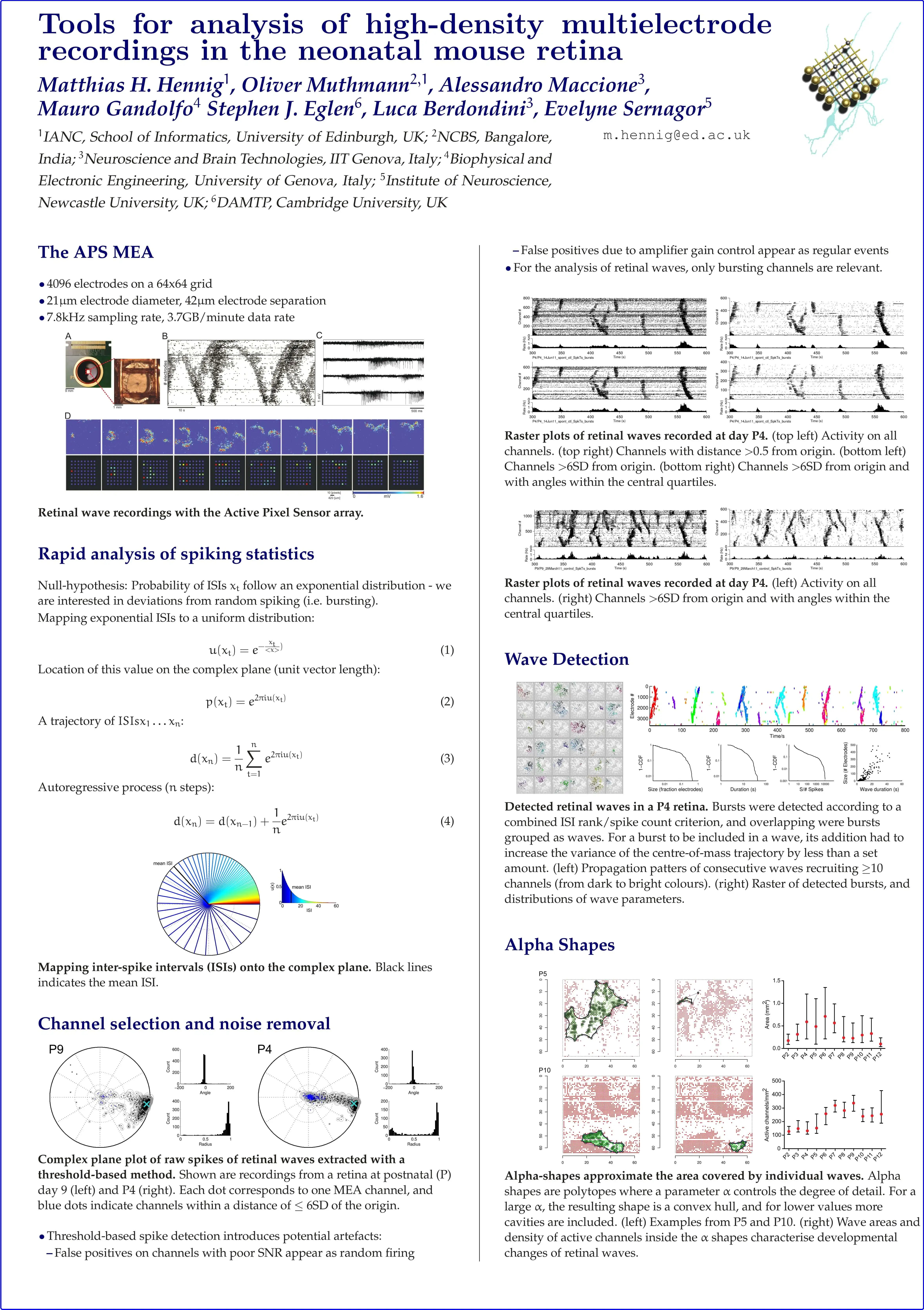
Tools for analysis of high-density multielectrode recordings in the neonatal mouse retina
Front. Neuroinform. (2013). DOI: 10.3389/conf.fninf.2013.08.00004
2013
Keywords:

Acute Retina
(conf. proc.)
Deciphering Retinal Functional Circuitry: High Resolution Large-Scale Population Recordings From The Mouse Retinal Ganglion Cell Layer
European Retina Meeting ERM (2013). Alicante, Spain.
2013
Keywords:

Brain Slices
(paper)
Dentate gyrus network dysfunctions precede the symptomatic phase in a genetic mouse model of seizures
Front. Cell. Neurosci. (2013). DOI: 10.3389/fncel.2013.00138.
2013
Keywords:
Neuronal circuit disturbances that lead to hyperexcitability in the cortico-hippocampal network are one of the landmarks of temporal lobe epilepsy. The dentate gyrus (DG) network plays an important role in regulating the excitability of the entire hippocampus by filtering and integrating information received via the perforant path. Here, we investigated possible epileptogenic abnormalities in the function of the DG neuronal network in the Synapsin II (Syn II) knockout mouse (Syn II−/−), a genetic mouse model of epilepsy. Syn II is a presynaptic protein whose deletion in mice reproducibly leads to generalized seizures starting at the age of 2 months. We made use of a high-resolution microelectrode array (4096 electrodes) and patch-clamp recordings, and found that in acute hippocampal slices of young pre-symptomatic (3–6 week-old) Syn II−/− mice excitatory synaptic output of the mossy fibers is reduced. Moreover, we showed that the main excitatory neurons present in the polymorphic layer of the DG, hilar mossy cells, display a reduced excitability. We also provide evidence of a predominantly inhibitory regulatory output from mossy cells to granule cells, through feed-forward inhibition, and show that the excitatory-inhibitory ratio is increased in both pre-symptomatic and symptomatic Syn II−/− mice. These results support the key role of the hilar mossy neurons in maintaining the normal excitability of the hippocampal network and show that the late epileptic phenotype of the Syn II−/− mice is preceded by neuronal circuitry dysfunctions. Our data provide new insights into the mechanisms of epileptogenesis in the Syn II−/− mice and open the possibility for early diagnosis and therapeutic interventions.

Neuronal Cultures
(conf. proc.)
Investigating the interplay between intrinsic and evoked activities in cultured neuronal networks by dimensional reduction techniques and high-density MEAs
BMC Neurosci.(2013). DOI: 10.1186/1471-2202-14-S1-P24.
2013
Keywords:
High density microelectrode arrays (MEAs) provide extracellular recordings from thousand of electrodes (http://www.3brain.com) and offer novel capabilities to investigate electrophysiological signaling in cultured neuronal networks and in ex vivo brain tissues. In this study we report on our recent technological and data analysis advancements to investigate the propagation and the interplay of spontaneous and electrically evoked activities in cultured networks.
Signal Processing
(conf. proc.)
Estimating the fraction of falsely detected spikes in high density microelectrode array recordings based on correlations
BMC Neurosci (2013). DOI: 10.1186/1471-2202-14-S1-P25
2013
Keywords:
High-density microelectrode arrays (MEA) can measure neuronal activity in potentially thousands of units with a high spatial resolution [1]. However due to the small size of the preamplifers, noise artifacts can affect spike detection. Additionally, the MEA chip itself is not perfectly homogeneous and the electrical coupling between the electrodes and a neuron may be weak. Therefore, the characteristics of neuronal spikes and noise are inherently different in each recording channel, such that estimating an average performance of the spike detection would not be representative for individual recording channels. As we aim to observe slow changes in single neuron activity, it is crucial to know how much a change in electrical coupling could potentially affect the number of detected spikes.
Signal Processing
(conf. proc.)
Homeostasis in large networks of neurons through the Ising model - do higher order interactions matter?
BMC Neurosci. (2013). DOI: 10.1186/1471-2202-14-S1-P166
2013
Keywords:
Homeostatic activity in large networks of neurons is a relatively scantly explored area of neuroscience, both on experimental and computational level [1]. With recent advance in recording techniques, the lack of experimental data is gradually ceasing to be the limitation. New multielectrode arrays (MEA) allow for monitoring cultures of thousands of neurons over many days with high spatial resolution [2]. However, the interpretation of multi-neuron recordings is not straightforward and requires methods going beyond the simplest descriptive statistics.

Technology
(conf. proc.)
Sensing and actuating electrophysiological activity on brain tissue and neuronal cultures with a high-density CMOS-MEA
The 17th International Conference on Solid-State Sensors, Actuators and Microsystems (TRANSDUCERS & EUROSENSORS XXVII) (2013). DOI: 10.1109/Transducers.2013.6626875.
2013
Keywords:
Multielectrode arrays (MEAs) for electrophysiological studies in neuroscience can nowadays be realized with Complementary Metal Oxide Semiconductor (CMOS) technology to provide large active areas with several thousands of simultaneously recording microelectrodes and electrode densities with inter-electrode separations approaching cellular sizes. This provides spatial and temporal resolutions to literally image electrophysiological activity propagations. Here, we focus on the achieved sensing capabilities of a 64×64 electrode array as well as on the actuation performances of a novel CMOS-chip integrating 16 electrodes for electrical stimulation. We report and discuss our recent results obtained from neuronal cell cultures, brain slices and mouse retina preparations.
.svg)


